You have no items in your shopping cart.
Description
Research Area
Signal Transduction
Images & Validation
−Item 1 of 3
| Tested Applications | IHC-P, WB |
|---|---|
| Dilution Range | WB - 1:1000, IHC-P - 1:50-100 |
| Reactivity | Human |
Key Properties
−| Host | Rabbit |
|---|---|
| Clonality | Polyclonal |
| Isotype | Rabbit IgG |
| Immunogen | This PKC-nu antibody is generated from rabbits immunized with a KLH conjugated synthetic peptide between 352-384 amino acids from human PKC-nu. Antigen Region: 352-384 aa. |
| Target | PRKD3 |
| Molecular Weight | 100471 Da |
| Conjugation | Unconjugated |
Storage & Handling
−| Storage | Maintain refrigerated at 2-8°C for up to 2 weeks. For long term storage store at -20°C in small aliquots to prevent freeze-thaw cycles |
|---|---|
| Form/Appearance | Purified polyclonal antibody supplied in PBS with 0.09% (W/V) sodium azide. This antibody is prepared by Saturated Ammonium Sulfate (SAS) precipitation followed by dialysis against PBS. |
| Expiration Date | 12 months from date of receipt. |
| Disclaimer | For research use only |
Alternative Names
−Serine/threonine-protein kinase D3, Protein kinase C nu type, Protein kinase EPK2, nPKC-nu, PRKD3, EPK2, PRKCN
Similar Products
−- Item 1 of 3
- Item 1 of 2
Phospho-PRKD3 (Ser41) Rabbit Polyclonal Antibody [orb4440]
IF, IHC-Fr, IHC-P
Bovine, Canine, Human, Mouse, Rabbit
Rat
Rabbit
Polyclonal
Unconjugated
100 μl, 50 μl, 200 μl - Item 1 of 1
- Item 1 of 1
- Item 1 of 1
PRKD3 Antibody [orb1316821]
IHC, WB
Human, Mouse, Rat
Mouse
Monoclonal
Unconjugated
100 μl, 50 μl, 30 μl

Quality Guarantee
Explore bioreagents carefree to elevate your research. All our products are rigorously tested for performance. If a product does not perform as described on its datasheet, our scientific support team will provide expert troubleshooting, a prompt replacement, or a refund. For full details, please see our Terms & Conditions and Buying Guide. Contact us at [email protected].
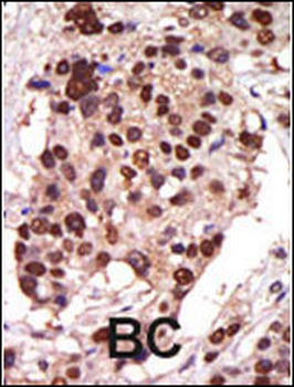
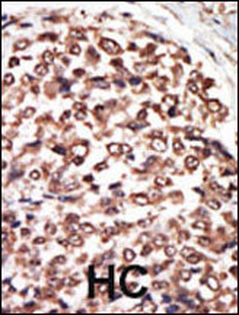
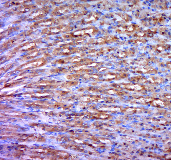
Formalin-fixed and paraffin-embedded human cancer tissue reacted with the primary antibody, which was peroxidase-conjugated to the secondary antibody, followed by AEC staining. This data demonstrates the use of this antibody for immunohistochemistry; clinical relevance has not been evaluated. BC = breast carcinoma; HC = hepatocarcinoma.
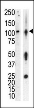
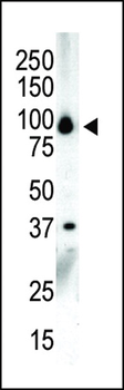
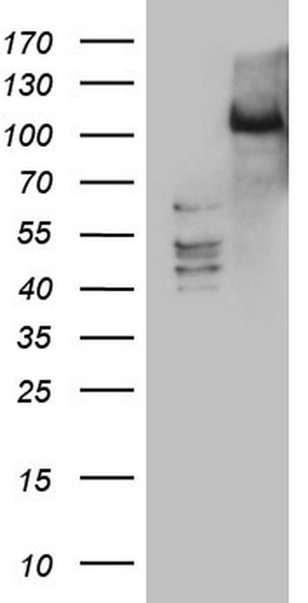
Western blot analysis of anti-PKCnu Pab in lysate of HL60 cells stimulated with PMA (lane A) and mouse brain tissue lysate (lane B). PKCnu (arrow) was detected using purified Pab. Secondary HRP-anti-rabbit was used for signal visualization with chemiluminescence.
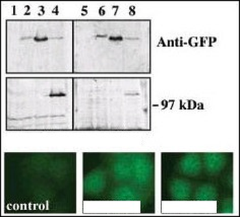
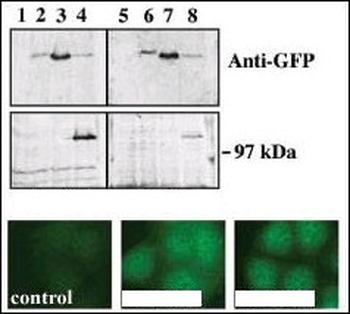
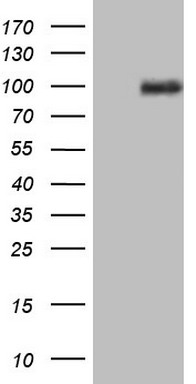
Upper panel, western blot analysis of GFP fusion protein expression in Panc-1 cells by using an anti-GFP antibody. Lanes 1 and 5: non-transfected cells; lanes 2 and 6: GFP-PKD-transfected cells; lanes 3 and 7: GFP-PKD2-transfected cells; lanes 4 and 8: GFP-PKD3 transfected cells. Center panel, western blot analysis of GFP fusion protein expression in Panc-1 cells by using PKD3 N-term and C-term antibodies. Lower panel, indirect immunofluorescence analysis of GFP-PKD3 fusion protein expression in Panc-1 cells by using antibodies.
Quick Database Links
UniProt Details
− No UniProt data available
NCBI Reference Sequences
−Associated Accession Numbers
Curated reference sequences for the gene transcript and protein product| Protein | NP_005804.1 |
|---|
Documents Download
Datasheet
Product Information
Request a Document
Protocol Information
WB
Western Blot (IB, immunoblot)
IHC-P
Immunohistochemistry Paraffin
PKC nu Antibody (orb1929637)

- 0.0
Based on 0 reviews
Participating in our Biorbyt product reviews program enables you to support fellow scientists by sharing your firsthand experience with our products.
Login to Submit a ReviewAvailable Sizes
Select a size below
Free Secondary Antibody (20 ul)0/0
Please add an antibody product to your cart first.