You have no items in your shopping cart.
Description
Research Area
Neuroscience
Images & Validation
−Item 1 of 5
| Tested Applications | ELISA, IF, IHC-P, WB |
|---|---|
| Reactivity | Human, Mouse, Rat |
| Application Notes |
Key Properties
−| Antibody Type | Primary Antibody |
|---|---|
| Host | Rabbit |
| Clonality | Polyclonal |
| Isotype | IgG |
| Immunogen | PION antibody was raised against a 19 amino acid synthetic peptide near the carboxy terminus of human PION.The immunogen is located within amino acids 770 - 820 of PION. |
| Target | PION |
| Purification | PION Antibody is affinity chromatography purified via peptide column. |
| Conjugation | Unconjugated |
Storage & Handling
−| Storage | Maintain refrigerated at 2-8°C for up to 2 weeks. For long term storage store at -20°C in small aliquots to prevent freeze-thaw cycles. |
|---|---|
| Form/Appearance | Liquid |
| Buffer/Preservatives | PION Antibody is supplied in PBS containing 0.02% sodium azide. |
| Concentration | 1 mg/mL |
| Expiration Date | 12 months from date of receipt. |
| Disclaimer | For research use only |
Alternative Names
−PION Antibody: PION, PION, Gamma-secretase-activating protein, Protein pigeon homolog, GSAP
Similar Products
−- Item 1 of 1
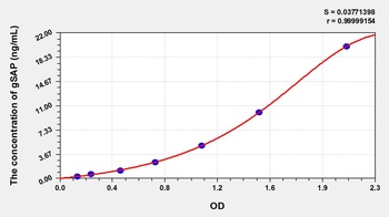
Rat Gamma-Secretase Activating Protein (gSAP) ELISA Kit [orb781862]
Rat
0.32-20 ng/mL
0.116 ng/mL
96 T, 48 T - Item 1 of 2
- Item 1 of 2
- Item 1 of 1
GSAP Rabbit Polyclonal Antibody [orb325416]
WB
Bovine, Canine, Equine, Rabbit, Rat
Human
Rabbit
Polyclonal
Unconjugated
100 μl 
GSAP Rabbit Polyclonal Antibody (Biotin) [orb2113933]
WB
Bovine, Canine, Equine, Human, Rabbit, Rat
Rabbit
Polyclonal
Biotin
100 μl

Quality Guarantee
Explore bioreagents carefree to elevate your research. All our products are rigorously tested for performance. If a product does not perform as described on its datasheet, our scientific support team will provide expert troubleshooting, a prompt replacement, or a refund. For full details, please see our Terms & Conditions and Buying Guide. Contact us at [email protected].
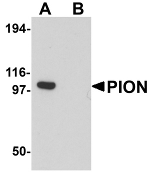
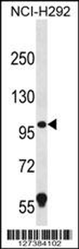
Western blot analysis of PION in EL4 cell lysate with PION antibody at 0.25 µg/mL in (A) the absence and (B) the presence of blocking peptide.
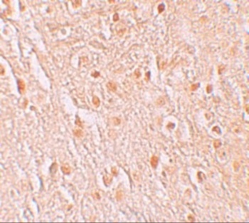
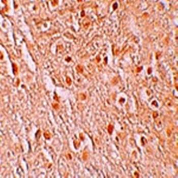
Immunohistochemistry of PION in human brain tissue with PION antibody at 5 µg/mL.
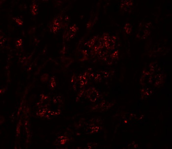
Immunofluorescence of PION in Human Brain cells with PION antibody at 20 µg/mL.
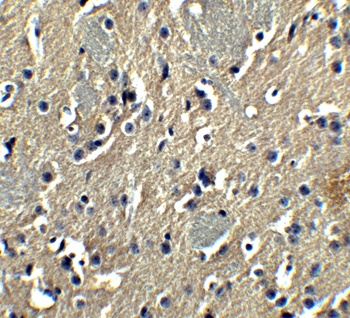
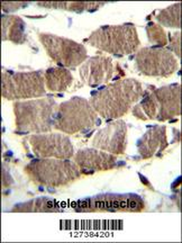
Immunohistochemistry of PION in mouse brain tissue with PION antibody at 5 µg/mL.
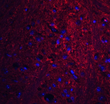
Immunofluorescence of PION in mouse brain tissue with PION antibody at 20 µg/mL. Red: PION Antibody (orb1239874) Blue: DAPI staining.
Documents Download
Datasheet
Product Information
Request a Document
Protocol Information
WB
Western Blot (IB, immunoblot)
IHC-P
Immunohistochemistry Paraffin
IF
Immunofluorescence
ELISA
Enzyme-linked Immunosorbent Assay (EIA)
PION Antibody (orb1239874)

- 0.0
Based on 0 reviews
Participating in our Biorbyt product reviews program enables you to support fellow scientists by sharing your firsthand experience with our products.
Login to Submit a ReviewAvailable Sizes
Select a size below
Free Secondary Antibody (20 ul)0/0
Please add an antibody product to your cart first.