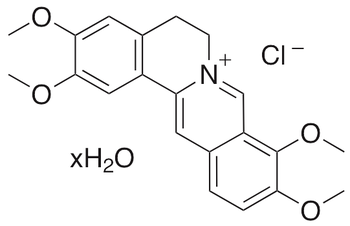
Palmatine is an inhibitor of dopamine generation. Palmatine could potentially be developed for the treatment of flavivirus infections. Palmatine has been used in the treatment of jaundice, dysentery, hypertension, inflammation, and liver-related diseases.(In Vitro):Palmatine (0-100 μM; 42 h) suppresses WNV with an EC50 value of 3.6 μM, and reduce the viral titers of DENV-2 and YFV with EC50 values of 26.4 μM and 7.3 μM, respectively.Palmatine (0-1128 μM; 24-72 h) inhibits colon cancer cell proliferation.Palmatine (0-704 μM; 24 h) reduces AURKA protein levels, induces G2/M phase arrest, and induces apoptosis in colon cancer cells via the mitochondrial associated pathway.(In Vivo):Palmatine (50 or 100 mg/kg; p.o.; daily for 7 days) ameliorates DSS (dextran sulfate sodium)-induced colitis and prevents infiltration of inflammatory cells.Palmatine (0-200 mg/kg; i.p.; once) attenuates D-galactosamine/Lipopolysaccharides (HY-D1056)-induced fulminant hepatic failure in mice.Palmatine (0-1 mg/kg; i.p.; 10 days) shows memory-enhancing activity in mice.Palmatine (33.75-135 mg/kg; p.o.; daily for 26 days) can effectively inhibit the growth of HCT-116 xenografts in mice.
Palmatine (50 or 100 mg/kg; p.o. ; daily for 7 days) ameliorates DSS (dextran sulfate sodium)-induced colitis and prevents infiltration of inflammatory cells. Palmatine (0-200 mg/kg; i.p.; once) attenuates D-galactosamine /Lipopolysaccharides (HY-D1056)-induced fulminant hepatic failure in mice. Palmatine (0-1 mg/kg; i.p.; 10 days) shows memory-enhancing activity in mice. Palmatine (33.75-135 mg/kg; p.o. ; daily for 26 days) can effectively inhibit the growth of HCT-116 xenografts in mice. Animal model: DSS-induced Colitis BALB/c mice model (8-week-old). Dosage: 50 or 100 mg/kg. Administration: Orally, daily, for 7 days. Result: Ameliorated DSS-induced colitis and prevented infiltration of inflammatory cells; remarkably extended the colon length; significantly suppressed the colonic MPO activity. Decreased the levels of colonic inflammatory cytokines (TNF-α, IFN-γ, IL-1β, IL-6, IL-4 and IL-10); Protected mucosal integrity by modulating TJs protein and apoptosis proteins; Restored DSS-induced decreases of TJ protein ZO-1, ZO-2 and claudin-1; Reduced Bax expression and enhanced Bcl-2 expression at the dose of 100 mg/kg, prevented epithelial apoptosis and improved intestinal integrity. Prevented DSS-induced changes of gut microbiota in colitis mice. Animal model: Male ICR mice (20–22 g), D-galactosamine/ lipopolysaccharide (GalN/LPS)-induced fulminant hepatic failure model. Dosage: 25, 50, 100, or 200 mg/kg. Administration: Intraperitoneal injection, 1 h before the GalN/LPS treatment. Result: Attenuated the mortality and serum aminotransferase activities increased by GalN/LPS. Prevented the increase of serum TNF-α and augmented that of serum IL-10. Decreased the TNF-a mRNA expression and increased the IL-10 mRNA expression. Attenuated the apoptosis of hepatocytes. Animal model: Swiss young male albino mice, with Scopolamine (HY-N0296)-and diazepam-induced amnesia model. Dosage: 0.1, 0.5, 1 mg/kg. Administration: Intraperitoneal injection, 10 days. Result: Significantly improved learning and memory of mice at 0.5 and 1 mg/kg and did not show any significant effect on locomotor activity of the mice. Significantly reversed scopolamine-and diazepam-induced amnesia in mice. Significantly reduced brain acetylcholinesterase activity of mice. Animal model: BALB/c-nude mice, HCT-116 xenograft model. Dosage: 33.75, 67.5 and 135 mg/kg. Administration: Oral administration, once a day for 26 days. Result: The tumor volume and weight of the treatment group were significantly reduced.
In Vitro
Palmatine (0-100 μM; 42 h) suppresses WNV with an EC50 value of 3.6 μM, and reduce the viral titers of DENV-2 and YFV with EC50 values of 26.4 μM and 7.3 μM, respectively. Palmatine (0-1128 μM; 24-72 h) inhibits colon cancer cell proliferation. Palmatine (0-704 μM; 24 h) reduces AURKA protein levels, induces G2/M phase arrest, and induces apoptosis in colon cancer cells via the mitochondrial associated pathway. Cell Proliferation Assay Cell line: HCT-116, SW480, HT-29. Concentration: 0, 88, 176, 352, and 704 μM (HCT-116, SW480); 0, 141, 282, 564, and 1128 μM (HT-29). Incubation time: 24, 48 and 72 h. Result: Decreased cell viability in a dose-dependent manner. Western blot analysis. Cell line: HCT-116, SW480, HT-29. Concentration: 100 nM for HCT-116, 500 nM for SW480 and HT-29. Incubation time: 24 h. Result: Promoted the expression of apoptosis markers such as P53 / P73, Caspase3, and Caspase9. Reduced AURKA protein levels. Increased cyt. c in the cytoplasm while reduced Bcl2 and Bcl-xl in a dose-dependent manner. Cell Cycle Analysis Cell line: HCT-116, SW480. Concentration: 88, 176, 352 and 704 μM. Incubation time: 24 h. Result: Induced G2/M phase arrest in a dose-dependent manner. Apoptosis Analysis Cell line: HCT-116, SW480. Concentration: 88, 176, 352 and 704 μM. Incubation time: 24 h. Result: Induced apoptosis in a dose-dependent manner.
Explore bioreagents carefree to elevate your research. All our products are rigorously tested for performance. If a product does not perform as described on its datasheet, our scientific support team will provide expert troubleshooting, a prompt replacement, or a refund. For full details, please see our Terms & Conditions and Buying Guide. Contact us at [email protected].


 Usually dispatched within 5-10 working days