You have no items in your shopping cart.
Description
Images & Validation
−Item 1 of 6
| Tested Applications | ELISA, IF, IHC, WB |
|---|---|
| Dilution Range | ELISA: 1:50,000-1:100,000, IHC: 1:200-1:600, IF: 1:5000, WB: 1:1000 - 1:5000 |
| Reactivity | Human |
| Application Notes |
Key Properties
−| Antibody Type | Primary Antibody |
|---|---|
| Host | Mouse |
| Clonality | Monoclonal |
| Isotype | IgG2a |
| Clone No. | 27F9.G4 |
| Immunogen | NFkB p65 (Rel A) peptide corresponding to a region near the C-terminus of the human protein conjugated to Keyhole Limpet Hemocyanin (KLH). |
| Target | RELA |
| Purity | This product was purified from concentrated tissue culture supernate by Protein A chromatography and showed a single band by IEP (immunoelectrophoresis) when tested with anti-mouse antibody. Reactivity was confirmed by ELISA against peptide conjugated carrier protein and by Western blot against HeLa whole cell lysate. |
| Conjugation | Unconjugated |
Storage & Handling
−| Storage | Store vial at -20° C or below prior to opening. This vial contains a relatively low volume of reagent (25 µL). To minimize loss of volume dilute 1:10 by adding 225 µL of the buffer stated above directly to the vial. Recap, mix thoroughly and briefly centrifuge to collect the volume at the bottom of the vial. Use this intermediate dilution when calculating final dilutions as recommended below. Store the vial at -20°C or below after dilution. Avoid cycles of freezing and thawing. |
|---|---|
| Form/Appearance | Liquid (sterile filtered) |
| Buffer/Preservatives | Preservative: 0.01% (w/v) Sodium Azide; Buffer: 0.02 M Potassium Phosphate, 0.15 M Sodium Chloride, pH 7.2 |
| Concentration | 0.98 mg/mL |
| Expiration Date | 12 months from date of receipt. |
| Dry Ice Shipping | Please note: This product requires shipment on dry ice. A dry ice surcharge will apply. |
| Disclaimer | For research use only |
Alternative Names
−mouse anti-NF-kB p65 Antibody, mouse anti-Rel A antibody, NFKB, nfkb, NF-kB, NF-kappaB, NFkappaB, Nuclear factor NF-kappa-B p65 subunit
Similar Products
−- Item 1 of 15
NFKB p65 Rabbit Polyclonal Antibody [orb11118]
FC, ICC
Bovine, Canine, Equine, Gallus, Porcine, Rabbit, Rat, Sheep, Zebrafish
Human, Mouse
Rabbit
Polyclonal
Unconjugated
50 μl, 100 μl, 200 μl - Item 1 of 9
NFKB p65 Rabbit Polyclonal Antibody [orb312399]
FC, ICC, IF, IHC-Fr, IHC-P, WB
Bovine, Canine, Porcine
Human, Mouse, Rat
Rabbit
Polyclonal
Unconjugated
50 μl, 100 μl, 200 μl - Item 1 of 10
NFKB p65 Mouse Monoclonal Antibody [orb500963]
ICC, IF, IHC-Fr, IHC-P, WB
Mouse, Rat
Human, Mouse, Rat
Mouse
Monoclonal
Unconjugated
200 μg, 50 μl, 100 μl, 200 μl - Item 1 of 8
Phospho-NFKB p65 (Ser468) Rabbit Polyclonal Antibody [orb6503]
FC, ICC, IF, IHC-Fr, IHC-P, WB
Bovine, Canine, Equine, Porcine
Human, Mouse, Rat
Rabbit
Polyclonal
Unconjugated
100 μl, 50 μl, 200 μl - Item 1 of 8
NFKB p65 Recombinant Rabbit Monoclonal Antibody [orb608066]
FC, ICC, IF, IHC-Fr, IHC-P, WB
Zebrafish
Human, Mouse, Rat
Rabbit
Recombinant
Unconjugated
50 μl, 100 μl, 25 μl


Quality Guarantee
Explore bioreagents carefree to elevate your research. All our products are rigorously tested for performance. If a product does not perform as described on its datasheet, our scientific support team will provide expert troubleshooting, a prompt replacement, or a refund. For full details, please see our Terms & Conditions and Buying Guide. Contact us at [email protected].
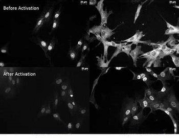
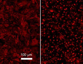
Anti NFkB monoclonal antibody – Immunocytochemistry. Tissue: Human Fibroblasts. Top: Before activation. Bottom: After activation with poly IC. Left: Anti-p65 NLS specific - (p/n orb345381). Right: Anti-p65 C-Term monoclonal antibody - (p/n orb344389). The two antibodies that are shown target different regions of the p65 protein. The different staining patterns are thought to correspond with different functional regions of the protein.
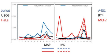
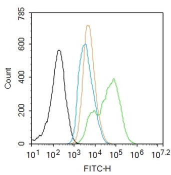
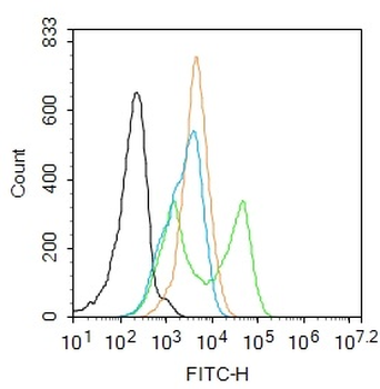
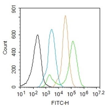
PAGE-MAP (microsphere affinity proteomics) of Mouse Anti-NFKB p65 (Rel A) Antibody. (orb344389). Antibody array western blot binding of gelfree size separated fractions of multiple lysates (solid lines) and shotgun mass spectroscopy identification (dashed lines) of the target band run in parallel correlate confirming the specificity of this antibody against NFKB p65.
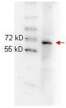
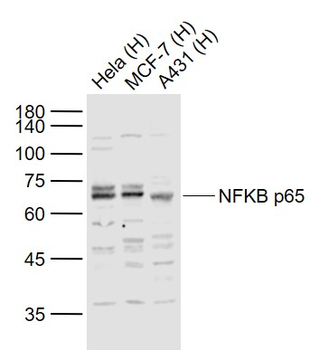
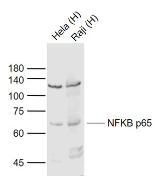
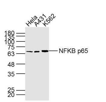
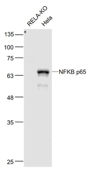
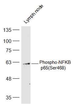
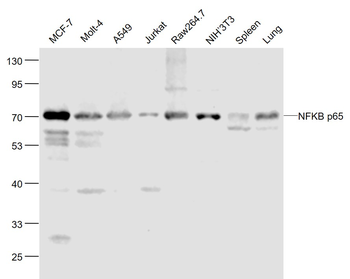
Biorbyt anti NFKB p65 (Rel A) monoclonal antibody (p/n orb344389) was used to detect ~65 kD band (red arrow) in HeLa whole cell lysate (p/n orb348668). Lysate was run on 4-20% gradient gel transferred under standard conditions and blocked in 1% BSA-TBST for 30 min at RT. Blot was probed with monoclonal anti-NFkB p65 at 1:1000 in 1% BSA-TBST o/n at 4°C and detected with HRP conjugated Rb-anti-Mouse antibody (p/n orb347544) at 1:40000 in (p/n orb348637) for 30 min at RT.
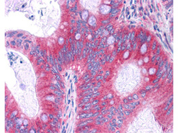
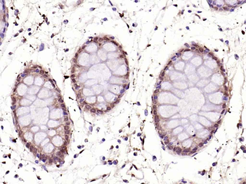
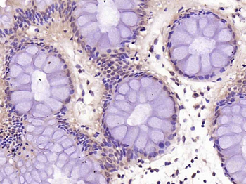
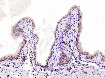
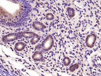
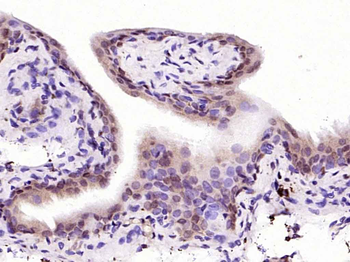
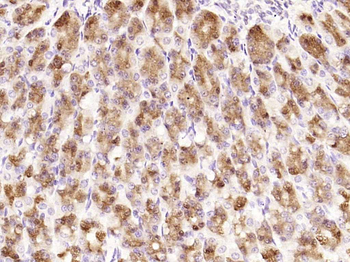
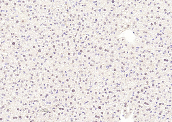
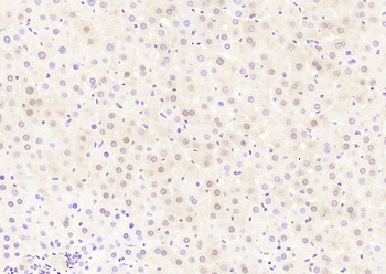
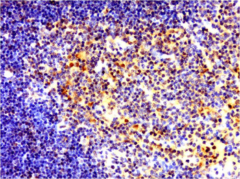
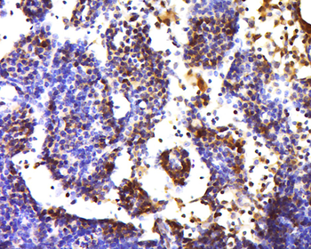
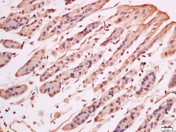
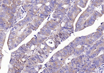
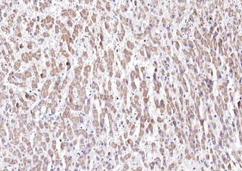
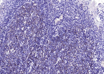
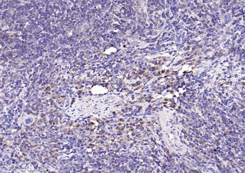
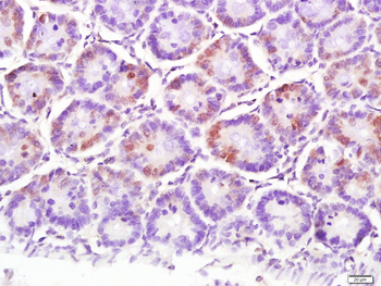
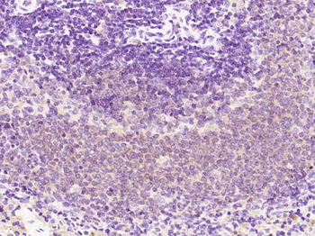
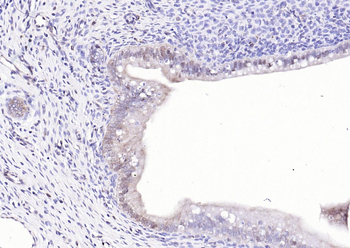
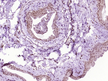
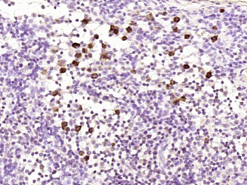
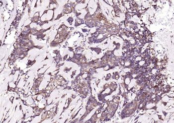
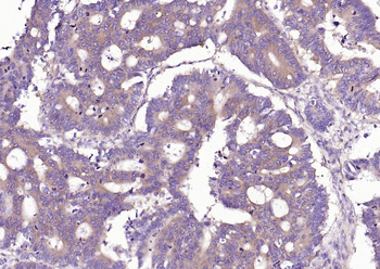
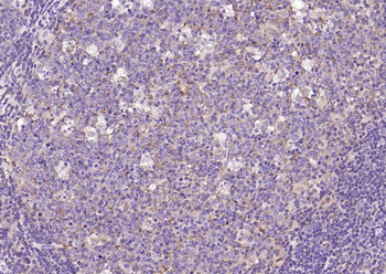
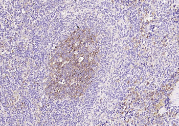
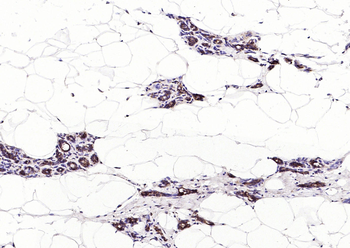
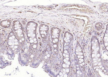
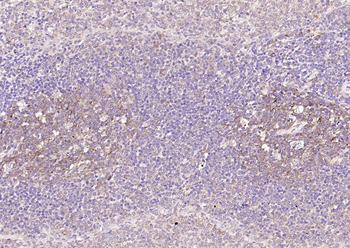
Biorbyt Antibody (p/n orb344389) has been tested in immunohistochemistry, analyzed by an anatomic pathologist and validated for use in IHC applications against formalin-fixed, paraffin-embedded human tissues. Showed moderate to strong staining within many tissues, including epithelium of the breast, small intestine, kidney, pancreas, prostate, skin, placenta, and uterus, as well as within neurons and lymphoid tissues such as spleen, thymus, and tonsil. The antibody produced an excellent signal with almost no background staining at a concentration of 2.5 µg/ml. The image displayed shows specific staining in colon carcinoma as the precipitated red signal, with a hematoxylin purple nuclear counterstain.
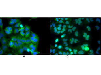
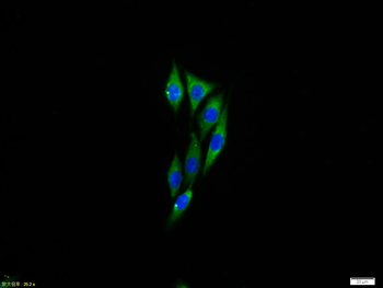
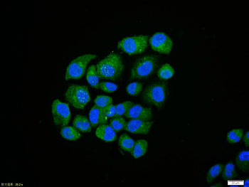
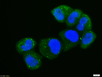
Biorbyt Monoclonal anti NFKB p65 (Rel A) antibody was used to detect p65 by immunofluorescence at a dilution of 1:5000. Hela cells were grown to sub-confluent on 18 mm2 glass coverslips #1.5. Cells were either unstimulated (A), or stimulated (B) with 50 ng/mL of TNF alpha for 30 min prior fixation. Cells were then fixed in methanol and blocked with 10% normal goat serum (NGS), in PBS, and TritonX 0.2% (Tx) and incubated for 1 hr at RT with primary ab, counterstained with DAPI and washed in PBS/NGS/Tx. Cells were incubated for 1 hr at RT with Atto 425 conjugated anti mouse secondary antibody for STED CW imaging.
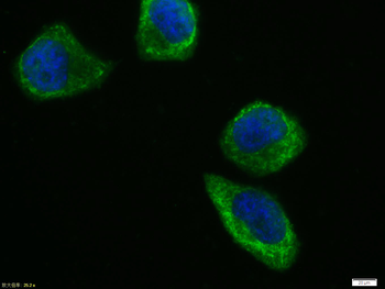
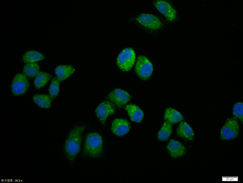
Biorbyt Monoclonal anti NFKB p65 (Rel A) antibody was used to detect p65 in b.end5 mouse endothelial cells. Unstimulated control cells (left) show cytoplasmic staining, TNF-alpha stimulated cells (right) show nuclear staining. For staining, cells were washed with PBS to remove all traces of culture media and fixed with paraformaldehyde 4% (45 min). Slides were washed with PBT (phosphate buffer 0.1M + Triton-X-100 0.1%), 3 times 10 min each, then, blocked with PBT + 5% normal goat serum, 1 hour. Sample was Incubated overnight in primary antibody (1:600 in blocking buffer). After 3X wash in PBT for 10 min, slides were incubated 1 hour with secondary antibody (1:1000) and mounted in 1:1 PB glycerol.
Documents Download
Datasheet
Product Information
Request a Document
Protocol Information
WB
Western Blot (IB, immunoblot)
IHC
Immunohistochemistry
IF
Immunofluorescence
ELISA
Enzyme-linked Immunosorbent Assay (EIA)
RELA Antibody (orb344390)

- 0.0
Based on 0 reviews
Participating in our Biorbyt product reviews program enables you to support fellow scientists by sharing your firsthand experience with our products.
Login to Submit a ReviewAvailable Sizes
Select a size below
Free Secondary Antibody (20 ul)0/0
Please add an antibody product to your cart first.