You have no items in your shopping cart.
Myogenin Antibody
SKU: orb385742
Description
Research Area
Developmental Biology, Epigenetics
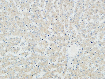
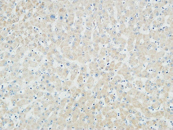
Images & Validation
−Item 1 of 1
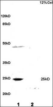
| Tested Applications | ELISA, IHC-P |
|---|---|
| Dilution Range | ELISA (order BSA/sodium azide-free format for coating),Immunohistochemistry (FFPE): 1-2ug/ml for 30 min at RT |
| Reactivity | Human, Mouse, Rat |
| Application Notes |
Key Properties
−| Antibody Type | Primary Antibody |
|---|---|
| Host | Mouse |
| Clonality | Monoclonal |
| Isotype | Mouse IgG1, kappa |
| Clone No. | F5D |
| Immunogen | Amino acids 138-158 from the rat protein were used as the immunogen for this Myogenin antibody. |
| Purification | Protein G affinity chromatography |
| Conjugation | Unconjugated |
Storage & Handling
−| Storage | Maintain refrigerated at 2-8°C for up to 2 weeks. For long term storage store at -20°C in small aliquots to prevent freeze-thaw cycles. |
|---|---|
| Buffer/Preservatives | 0.2 mg/ml in 1X PBS with 0.1 mg/ml rAlbumin and 0.05% sodium azide |
| Expiration Date | 12 months from date of receipt. |
| Disclaimer | For research use only |
Similar Products
−- Item 1 of 2
Myogenin Rabbit Polyclonal Antibody [orb6492]
WB
Bovine, Canine, Equine, Guinea pig, Human, Porcine, Rabbit, Sheep
Mouse, Rat
Rabbit
Polyclonal
Unconjugated
50 μl, 100 μl, 200 μl - Item 1 of 4
Myogenin rabbit pAb Antibody [orb765753]
ELISA, IF, IHC, WB
Human, Mouse, Porcine, Rat
Polyclonal
Unconjugated
100 μl - Item 1 of 4
MYOG Antibody [orb1410087]
ELISA, FC, IF, WB
Human, Mouse, Rat
Mouse
Monoclonal
Unconjugated
20 μg, 100 μg, 100 μg (without BSA and Azide) - Item 1 of 6
Alpha Smooth Muscle Actin Antibody [orb749771]
IF, IHC-P
Human, Rat
Mouse
Monoclonal
Unconjugated
100 μg, 20 μg - Item 1 of 6
ACTA2 Antibody Cocktail / Alpha Actin [orb749772]
IF, IHC-P
Human, Rat
Mouse
Monoclonal
Unconjugated
100 μg, 20 μg


Quality Guarantee
Explore bioreagents carefree to elevate your research. All our products are rigorously tested for performance. If a product does not perform as described on its datasheet, our scientific support team will provide expert troubleshooting, a prompt replacement, or a refund. For full details, please see our Terms & Conditions and Buying Guide. Contact us at [email protected].
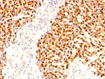
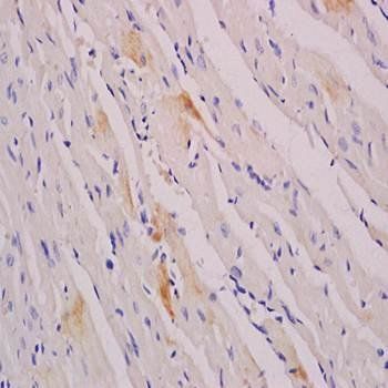
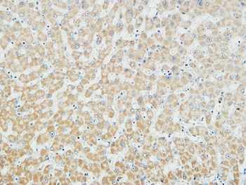
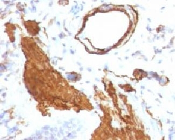
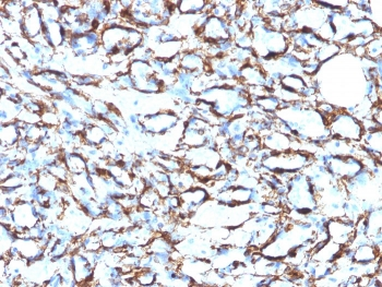
IHC: Formalin-fixed, paraffin-embedded human Rhabdomyosarcoma stained with Myogenin antibody (F5D)
Quick Database Links
UniProt
UniProt Details
− No UniProt data available
Documents Download
Datasheet
Product Information
Request a Document
Protocol Information
IHC-P
Immunohistochemistry Paraffin
ELISA
Enzyme-linked Immunosorbent Assay (EIA)
Myogenin Antibody (orb385742)

- 0.0
Based on 0 reviews
Participating in our Biorbyt product reviews program enables you to support fellow scientists by sharing your firsthand experience with our products.
Login to Submit a ReviewAvailable Sizes
Select a size below