You have no items in your shopping cart.
Description
Research Area
Immunology & Inflammation
Images & Validation
−Item 1 of 5
| Tested Applications | ELISA, IHC-P, WB |
|---|---|
| Dilution Range | Western blot: 1-2ug/ml,Immunohistochemistry (FFPE): 0.5-1ug/ml for 30 min at RT,ELISA: order the BSA free format for coating |
| Reactivity | Human |
| Application Notes |
Key Properties
−| Antibody Type | Primary Antibody |
|---|---|
| Host | Mouse |
| Clonality | Monoclonal |
| Isotype | Mouse IgG1, kappa |
| Clone No. | 139H2 |
| Immunogen | Human milk-fat globule membranes (HMFGM) was used as the immunogen for the MUC1 antibody. |
| Purification | Protein G affinity chromatography |
| Conjugation | Unconjugated |
Storage & Handling
−| Storage | Maintain refrigerated at 2-8°C for up to 2 weeks. For long term storage store at -20°C in small aliquots to prevent freeze-thaw cycles. |
|---|---|
| Buffer/Preservatives | 0.2 mg/ml in 1X PBS with 0.1 mg/ml rAlbumin and 0.05% sodium azide |
| Expiration Date | 12 months from date of receipt. |
| Disclaimer | For research use only |
Similar Products
−- Item 1 of 11
MUC1 Rabbit Polyclonal Antibody [orb4746]
ICC, IF, IHC-Fr, IHC-P
Mouse, Rat
Human, Mouse, Rat
Rabbit
Polyclonal
Unconjugated
50 μl, 100 μl, 200 μl - Item 1 of 7
MUC1 Rabbit Polyclonal Antibody [orb315622]
ICC, IF, IHC-P, WB
Human, Porcine, Rat
Rabbit
Polyclonal
Unconjugated
100 μg - Item 1 of 7
MUC1 Rabbit Polyclonal Antibody [orb578068]
IHC, WB
Bovine, Canine, Equine, Guinea pig, Rabbit, Rat
Human, Mouse, Porcine
Rabbit
Polyclonal
Unconjugated
100 μl - Item 1 of 8
- Item 1 of 6
MUC1 Antibody [orb388454]
IHC, WB
Human, Mouse
Mouse
Monoclonal
Unconjugated
20 μg, 100 μg, 100 μg (without BSA and Azide)


Quality Guarantee
Explore bioreagents carefree to elevate your research. All our products are rigorously tested for performance. If a product does not perform as described on its datasheet, our scientific support team will provide expert troubleshooting, a prompt replacement, or a refund. For full details, please see our Terms & Conditions and Buying Guide. Contact us at [email protected].
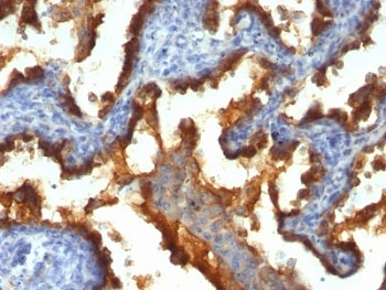
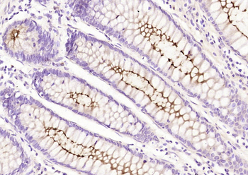
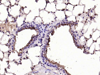
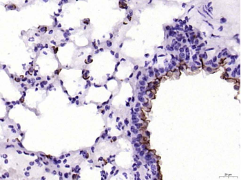
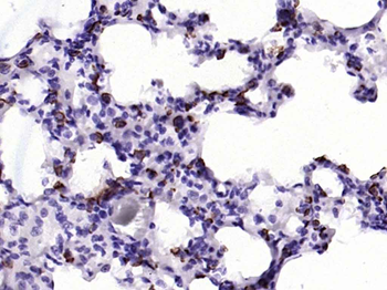
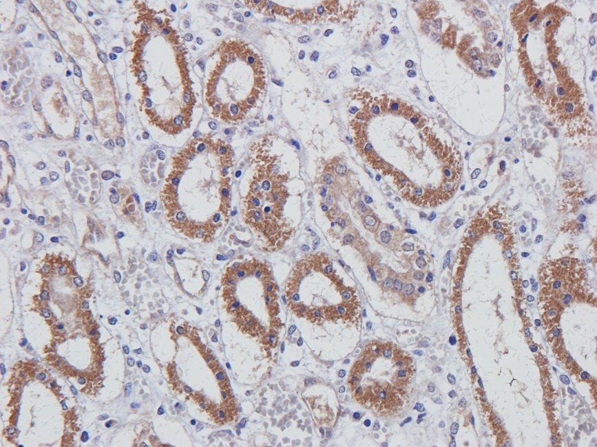
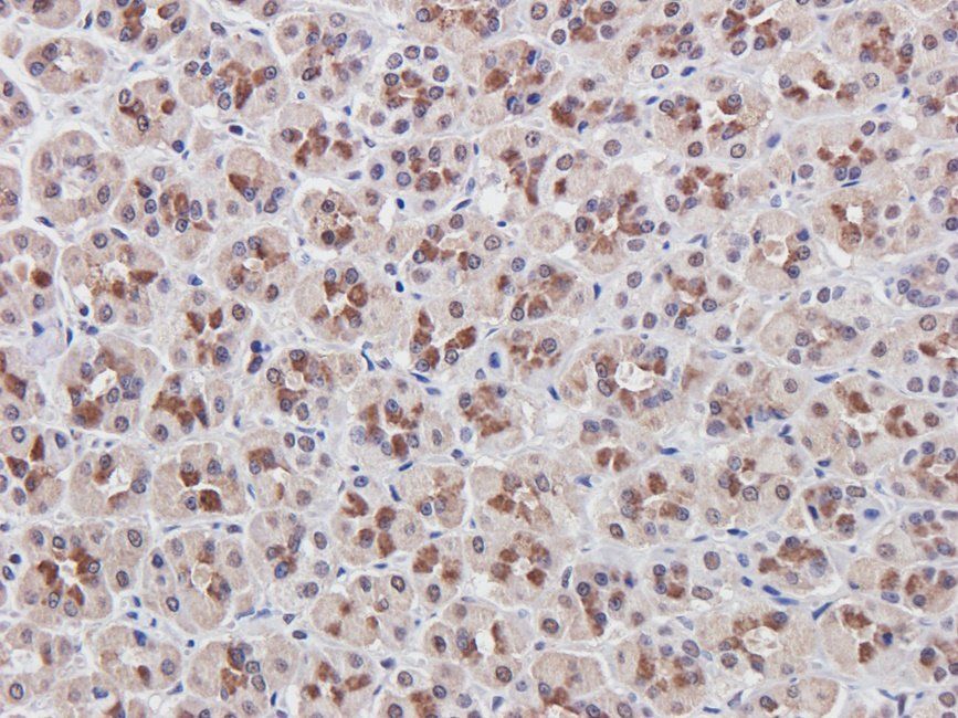
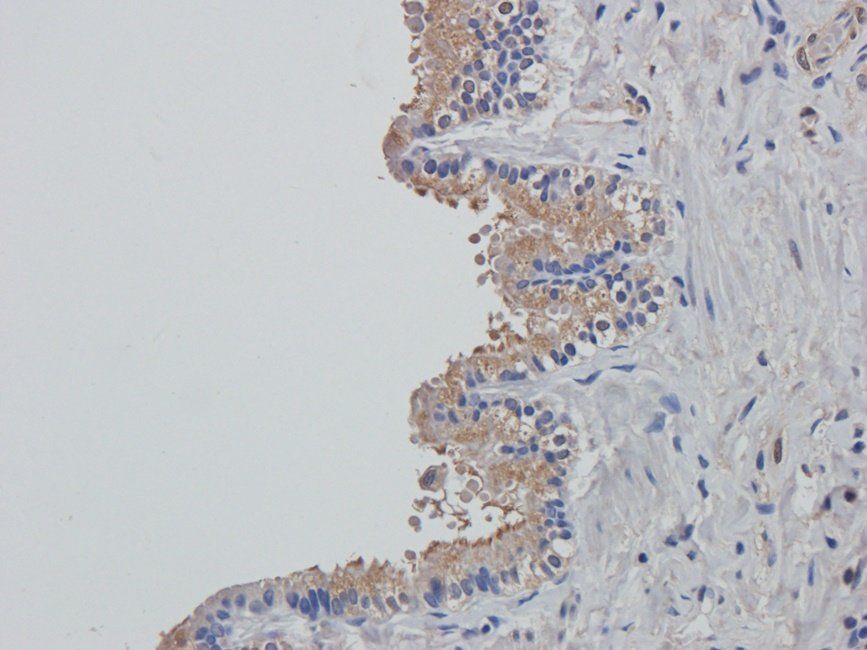
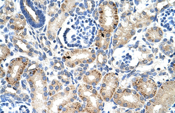
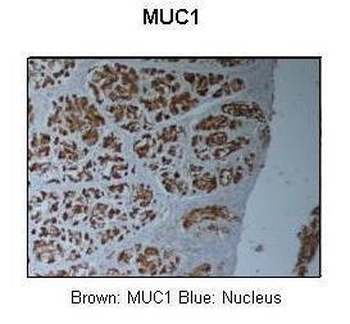
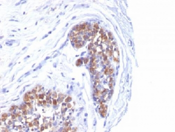
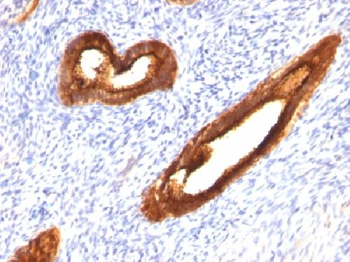
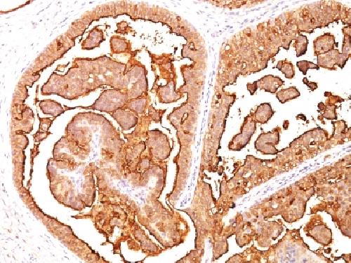
IHC: Formalin-fixed, paraffin-embedded human lung cancer stained with MUC1 antibody (clone 139H2).
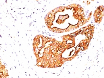
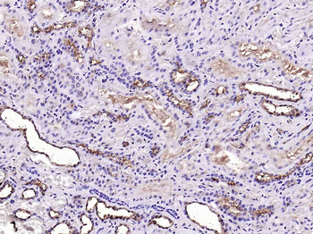
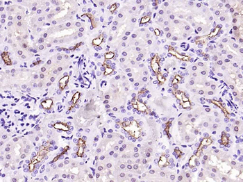
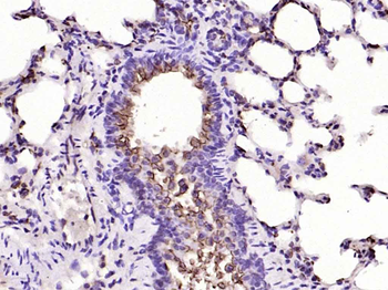
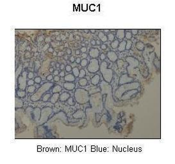
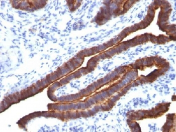
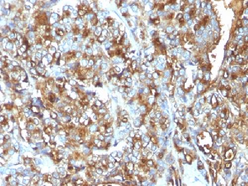
IHC: Formalin-fixed, paraffin-embedded human breast cancer stained with MUC1 antibody (clone 139H2).
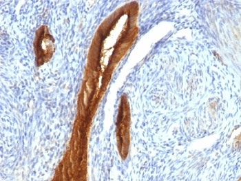
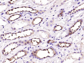
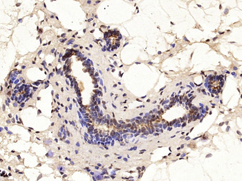
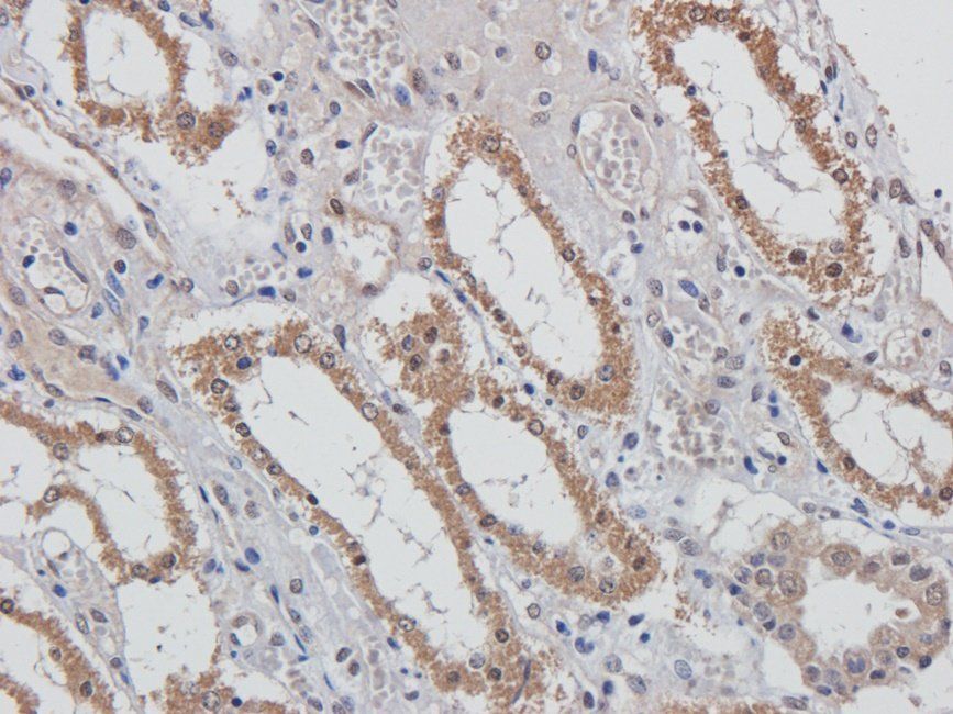
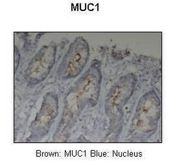
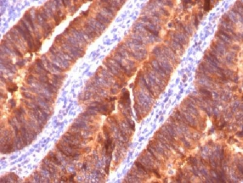
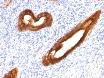
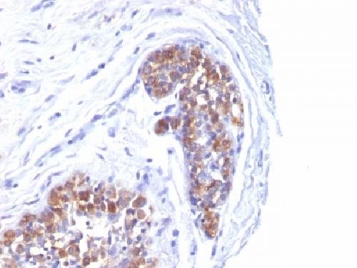
IHC: Formalin-fixed, paraffin-embedded human endometrial carcinoma stained with MUC1 antibody (clone 139H2).
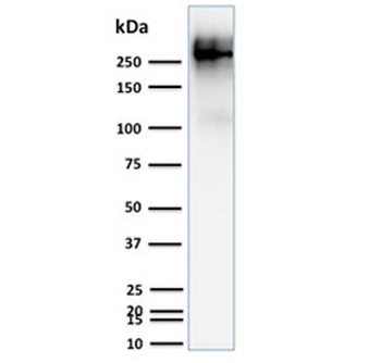
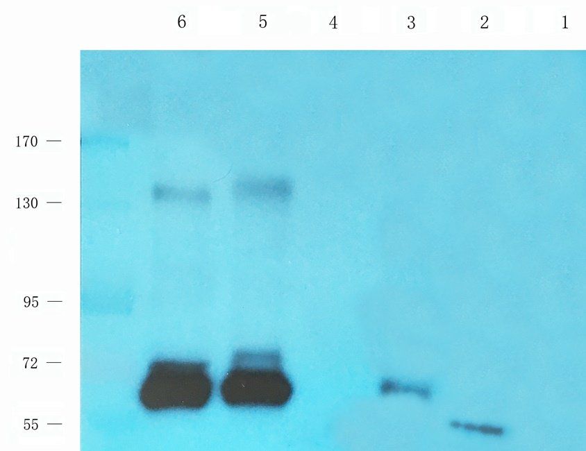
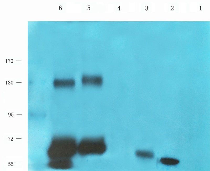
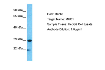
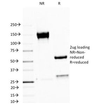
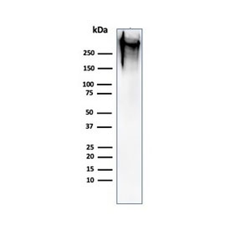
Western blot testing of human T47D cell lysate with MUC1 antibody. This glycoprotein is commonly visualized between 120~500 kDa.
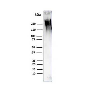
Western blot testing of human MCF7 cell lysate with MUC1 antibody. This glycoprotein is commonly visualized between 120~500 kDa.
Quick Database Links
UniProt
UniProt Details
− No UniProt data available
Documents Download
Datasheet
Product Information
Request a Document
Protocol Information
WB
Western Blot (IB, immunoblot)
IHC-P
Immunohistochemistry Paraffin
ELISA
Enzyme-linked Immunosorbent Assay (EIA)
MUC1 Antibody (orb749682)

- 0.0
Based on 0 reviews
Participating in our Biorbyt product reviews program enables you to support fellow scientists by sharing your firsthand experience with our products.
Login to Submit a ReviewAvailable Sizes
Select a size below
Choose Conjugation or Carrier Free Version
Free Secondary Antibody (20 ul)0/0
Please add an antibody product to your cart first.