
You have no items in your shopping cart.
Fluorescent Protein Research
mRuby: A Bright, Monomeric Red Fluorescent Protein
Explore the unique large Stokes shift and high brightness of mRuby, compare its performance to mCherry, and discover reagents for detection and FRET biosensor development.
1. What Is mRuby?
mRuby is an engineered, highly bright, monomeric red fluorescent protein (RFP). It was originally developed through the directed evolution of eqFP611, a tetrameric red fluorescent protein isolated from the sea anemone Entacmaea quadricolor.
First published by Kredel et al. in 2009, mRuby was designed to overcome the limitations of early RFPs, such as poor maturation and oligomerization, which disrupted fusion protein functions. With an extinction coefficient of ~112,000 M⁻¹cm⁻¹ and a quantum yield of 0.35, mRuby stands out as one of the brightest first-generation monomeric RFPs available to researchers.
Key Advantage: mRuby features a uniquely large Stokes shift (~47 nm). This wide gap between excitation (558 nm) and emission (605 nm) drastically reduces background signal from excitation light scattering, making it exceptional for deep-tissue imaging and FRET.
mRuby at a Glance
Property | mRuby |
|---|---|
Origin species | Entacmaea quadricolor (Sea Anemone) |
Excitation Max | 558 nm |
Emission Max | 605 nm |
Stokes Shift | 47 nm (Exceptionally large) |
Extinction Coefficient | 112,000 M⁻¹cm⁻¹ |
Quantum Yield | 0.35 |
Quaternary Structure | Monomer |
2. mRuby vs. mCherry
When choosing a red fluorescent protein, mCherry (derived from Discosoma sp.) is historically the most common choice due to its incredibly fast maturation and high photostability. However, mRuby is significantly brighter and offers spectral advantages for certain experiments.
Feature | mRuby | mCherry |
|---|---|---|
Overall Brightness | High (approx. 2.5x brighter than mCherry) | Moderate |
Stokes Shift | 47 nm (Great for reducing bleed-through) | 23 nm (Standard) |
Maturation Rate | Moderate (requires more time to fully mature) | Extremely fast |
Photostability | Moderate | High |
Selection guidance: Choose mRuby when absolute brightness is the limiting factor in your assay or when designing a FRET biosensor that benefits from a large Stokes shift. Choose mCherry if you need ultra-fast reporting of rapid gene expression events.
3. Key Research Applications
Multicolor Imaging
mRuby pairs beautifully with green fluorescent proteins like GFP, EGFP, or mNeonGreen. Its distinct excitation and emission spectra ensure clean separation across fluorescent channels with minimal crosstalk.
FRET Biosensors
Because of its large extinction coefficient and Stokes shift, mRuby is highly effective as a FRET acceptor when paired with a green donor (e.g., Clover-mRuby pairs), enabling sensitive live-cell tracking of molecular interactions.
Protein Fusions
As a strict monomer, mRuby can be successfully fused to structural proteins (like actin or tubulin) without causing the unwanted oligomerization or aggregation seen in older tetrameric red fluorescent proteins.
4. The Evolution of the mRuby Family
Since its introduction in 2009, mRuby has served as the foundational scaffold for subsequent generations of enhanced red fluorescent proteins. While standard mRuby remains a powerful tool, researchers have successfully engineered optimized variants:
- mRuby2 (2012): Engineered for improved photostability and higher folding efficiency, specifically tailored to enhance the performance of green/red FRET biosensors.
- mRuby3 (2016): Further optimized for extreme brightness and photostability, competing directly with the brightest modern RFPs for demanding applications like super-resolution imaging.
Note: Antibodies raised against the original mRuby protein generally cross-react excellently with mRuby2 and mRuby3 due to high sequence conservation across the variants.
5. Recommended mRuby Antibodies
To reliably detect mRuby fusion proteins in non-fluorescent assays, specific anti-mRuby antibodies are required. Because mRuby is derived from the sea anemone rather than jellyfish or coral, antibodies targeting GFP or DsRed will typically fail to recognize it.
Featured Product for mRuby Detection
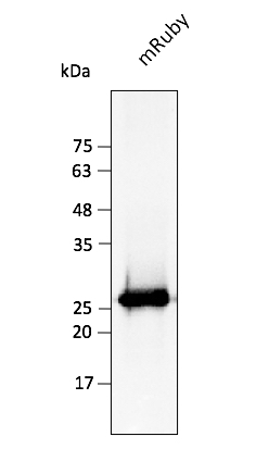
mRuby Antibody orb1463286.
Highly Specific Recognition
mRuby Antibody — orb1463286
orb1463286 is a robust antibody explicitly raised to detect mRuby. It provides high sensitivity for confirming the expression and molecular weight of mRuby fusion proteins in your experimental models. This reagent is essential for Western Blot validation where direct fluorescence is lost due to sample denaturation.
Target: mRuby
Application: WB (Western Blot)
Reactivity: Entacmaea quadricolor derivatives
Conjugation: Unconjugated
6. Detecting and Validating mRuby Constructs
Direct Fluorescence
mRuby can be imaged using standard TRITC or Cy3 filter sets. Due to its large Stokes shift, researchers can utilize excitation filters near 558 nm and collect emission safely above 600 nm, dramatically reducing scattered excitation light and cellular autofluorescence.
Antibody Detection
Direct fluorescence confirms the fluorophore has properly folded, but it does not confirm the fusion protein remains completely intact. Utilize the targeted orb1463286 antibody in Western Blots to ensure your mRuby-fusion protein is fully expressed and has not undergone unexpected proteolytic cleavage.
7. Frequently Asked Questions
Can I use an anti-DsRed or anti-mCherry antibody to detect mRuby?
Generally, no. mRuby is derived from Entacmaea quadricolor (eqFP611), while DsRed and mCherry are derived from Discosoma sp. Due to distinct evolutionary origins and structural differences, you should use an antibody specifically raised against mRuby.
Is mRuby safe to use for fusion proteins?
Yes. Kredel et al. extensively engineered mRuby to break the tetrameric interfaces of its wild-type parent. mRuby behaves as a strict monomer in live cells, preventing the artificial aggregation often seen when using older red fluorescent proteins as fusion tags.
Why is a "Large Stokes Shift" important?
The Stokes shift is the distance (in nanometers) between the peak excitation wavelength and the peak emission wavelength. mRuby's large 47 nm shift means the emitted fluorescence is spectrally far away from the excitation laser/light, making it much easier for microscope filters to block out background noise and deliver a cleaner, high-contrast image.
8. Scientific References
- Kredel S, et al. mRuby, a bright monomeric red fluorescent protein for labeling of subcellular structures. PLOS One. 2009;4(2):e4391. doi:10.1371/journal.pone.0004391
- Lam AJ, et al. Improving FRET dynamic range with bright green and red fluorescent proteins. Nature Methods. 2012;9(10):1005-1012. doi:10.1038/nmeth.2171 (Development of mRuby2)
- Bajar BT, et al. Improving brightness and photostability of green and red fluorescent proteins for live cell imaging and FRET reporting. Nature Communications. 2016;7:11305. doi:10.1038/ncomms11305 (Development of mRuby3)
- Lee S, et al. A highly improved red fluorescent protein for FRET/FLIM measurements. Scientific Reports. 2016;6:20889. doi:10.1038/srep20889