
You have no items in your shopping cart.
Description
Research Area
Protein Biochemistry
Images & Validation
−Item 1 of 3
| Tested Applications | SDS-PAGE |
|---|---|
| Application Notes |
Key Properties
−| Source | Mouse |
|---|---|
| Biological Origin | Mouse |
| Isotype | IgG F(c) |
| Conjugation | Unconjugated |
| Purity | MOUSE IgG F(c) fragment was prepared from normal serum by a multi-step process which includes delipidation, salt fractionation, ion exchange chromatography and papain digestion followed by chromatographic separation and extensive dialysis against the buffer stated above. Assay by immunoelectrophoresis resulted in a single precipitin arc against anti-Mouse Serum, anti-Mouse IgG and anti-Mouse IgG F(c). No reaction was observed against anti-Mouse IgG F(ab’)2 or anti-Papain. |
Storage & Handling
−| Storage | Store vial at 4° C prior to opening. For extended storage aliquot contents and freeze at -20° C or below. Avoid cycles of freezing and thawing. Centrifuge product if not completely clear after standing at room temperature. Mouse IgG Fc fragment is stable for several weeks at 4° C as an undiluted liquid. Dilute only prior to immediate use. |
|---|---|
| Form/Appearance | Liquid (sterile filtered) |
| Buffer/Preservatives | Preservative: 0.01% (w/v) Sodium Azide; Buffer: 0.02 M Potassium Phosphate, 0.15 M Sodium Chloride, pH 7.2 |
| Concentration | 1.1 mg/mL |
| Expiration Date | 12 months from date of receipt. |
| Disclaimer | For research use only |
Alternative Names
−MOUSE IgG F(c) fragment, Mouse Fc, Immunoglobulin Fc
Similar Products
−- Item 1 of 3
Mouse IgG Fc Antibody Alkaline Phosphatase Conjugated [orb347559]
DOT, ELISA, IHC, WB
Mouse
Rabbit
Polyclonal
AP
1 mg - Item 1 of 2
Mouse IgG F(c) Biotin Antibody [orb346295]
DOT
Biotin
This product was prepared from normal serum delipidation, salt fractionation, ion exchange chromatography followed by papain digestion and extensive dialysis against the buffer stated above. Assay by immunoelectrophoresis resulted in a single precipitin arc against anti-biotin, anti-Mouse IgG, anti-Mouse IgG F(c) and anti-Mouse Serum. No reaction was observed against anti-Mouse IgG F(ab’)2 or anti-Papain.
Mouse
1 mg - Item 1 of 2
Mouse IgG Fc Antibody [orb750706]
ELISA, IHC, KO/KD Validated, WB
Mouse
Goat
Polyclonal
Unconjugated
2 ml - Item 1 of 1
- Item 1 of 1


Quality Guarantee
Explore bioreagents carefree to elevate your research. All our products are rigorously tested for performance. If a product does not perform as described on its datasheet, our scientific support team will provide expert troubleshooting, a prompt replacement, or a refund. For full details, please see our Terms & Conditions and Buying Guide. Contact us at [email protected].
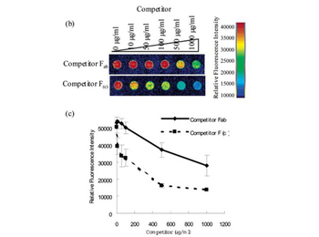
Ability of ProteoChip to bind the antibody F(c) region. (b) Scanning images of protein microarray: competition between FITC-labeled F(c) [p/n orb346285] and unlabeled Fab fragments [p/n orb346282] (upper picture), and FITC-labeled F(c) [p/n orb346285] and unlabeled F(c) fragments [orb346280] (lower picture). (c) Scanning images were analyzed using QuantumArray software, and fluorescence intensities of each spot were plotted versus competitor concentration. Competition between FITC-labeled F(c) and unlabeled Fab fragment, and FITC-labeled F(c) and unlabeled F(c) fragments, are shown by the solid line and broken line, respectively.
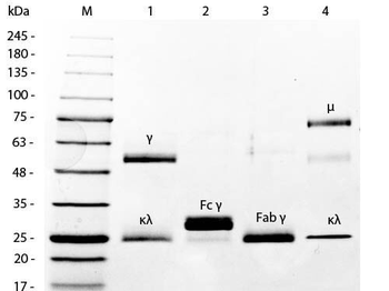
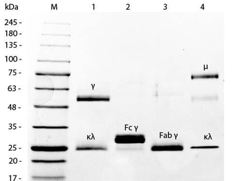
SDS-PAGE of Mouse IgG Whole Molecule Rhodamine Conjugated (p/n orb346272). MW: 5 µl Opal Prestained Marker. Lane 1: Reduced Mouse IgG Whole Molecule Rhodamine Conjugated (p/n orb346272). Lane 2: Reduced Mouse F(c) Fragment (p/n orb346280). Lane 3: Reduced Mouse F(ab) Fragment (p/n orb346282). Lane 4: Mouse IgM Kappa Myeloma Protein. Load: 1 µg per lane. Predicted/Observed size: IgG at 50 and 25 kDa; F(c) at 25 kDa; F(ab) at 25 kDa; IgM K at 70 and 23 kDa. Observed F(c) Fragment migrates slightly higher.
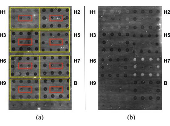
Strong responses to polyclonal anti-HA antiserum are readily observable on an AIR hemagglutinin microarray. (a) 1% BSA control. (b) Anti-H7 polyclonal antiserum (A/Netherlands/219/2003, H7N7), 1:80 dilution (1.3%) in 1% BSA. Spots showing substantially increased brightness indicate binding to immobilized H7. In both cases, antigens were arrayed in square patterns as indicated by the yellow boxes in (a); a mouse IgG Fc domain (p/n orb346280) was included as negative control (red boxes). Slight differences in spot intensity in the control (a) are due to differences in deposition efficiency for different antigens or controls. Specific antigens used in these experiments are indicated in Table 2. Goat anti-fluorescein, (p/n orb345272) used as an internal negative control.
Documents Download
Datasheet
Product Information
Request a Document
Protocol Information
Protein Handling and Storage Guide
Protein Handling Guide
SDS-PAGE
Sodium Dodecyl Sulphate PolyAcrylamide Gel Electrophoresis
Mouse IgG F(c) Antibody (orb346280)

- 0.0
Based on 0 reviews
Participating in our Biorbyt product reviews program enables you to support fellow scientists by sharing your firsthand experience with our products.
Login to Submit a ReviewAvailable Sizes
Select a size below
 Usually dispatched within 5-10 working days