
You have no items in your shopping cart.
Fluorescent Protein Research
mNeonGreen: The Brightest Monomeric Yellow-Green Fluorescent Protein
Explore the extreme brightness and photostability of mNeonGreen, compare its performance to EGFP and mClover3, and discover the split-mNG2 system for CRISPR-based endogenous protein tagging.
1. What Is mNeonGreen?
Unlike traditional green fluorescent proteins derived from Aequorea victoria jellyfish, mNeonGreen is an engineered monomeric yellow-green fluorescent protein derived from a tetrameric protein found in the lancelet (cephalochordate) Branchiostoma lanceolatum.
First characterized in 2013 by Shaner et al., it was explicitly developed to push the limits of fluorescence intensity and photostability. Boasting a quantum yield of 0.80 and an exceptionally high extinction coefficient (116,000 M⁻¹cm⁻¹), mNeonGreen is widely recognized as the brightest monomeric green or yellow fluorescent protein currently available.
Key point: mNeonGreen is exceptionally bright and photostable, making it a premier choice for demanding applications like stochastic single-molecule localization microscopy (SMLM) and low-abundance endogenous protein tracking.
mNeonGreen at a Glance
Property | mNeonGreen |
|---|---|
Origin species | Branchiostoma lanceolatum (Lancelet) |
Excitation Max | 506 nm |
Emission Max | 517 nm |
Extinction Coefficient | 116,000 M⁻¹cm⁻¹ |
Quantum Yield | 0.80 |
pKa | 5.7 (Excellent acid tolerance) |
Maturation Time | Exceptionally fast (~3-fold faster than mEGFP) |
2. mNeonGreen vs EGFP and Clover
While EGFP remains a highly utilized general-purpose reporter, it often falls short in modern, photon-hungry super-resolution techniques. mClover3 offers improved brightness over EGFP but can suffer from faster photobleaching. mNeonGreen bridges these gaps by offering unparalleled brightness without sacrificing stability.
Feature | mNeonGreen | EGFP | mClover3 |
|---|---|---|---|
In Vivo Brightness | ~3- to 5-fold brighter than EGFP | Baseline reference | Similar to mNeonGreen |
Photostability | Superior (~3-fold higher than Clover) | High | Low/Moderate |
Monomeric Character | Strict monomer (ideal for fusions) | Weak dimer at high concentrations | Strict monomer |
Acid Tolerance | High (pKa 5.7) | Moderate (pKa ~6.0) | Low (pKa 6.2) |
Selection guidance: Switch from EGFP to mNeonGreen when imaging low-abundance proteins or utilizing single-molecule super-resolution microscopy. Switch from Clover to mNeonGreen if your construct suffers from rapid photobleaching under sustained laser illumination.
3. Key Research Applications
Super-Resolution Imaging
With an exceptional photon yield (~660 photons per localized molecule), mNeonGreen outperforms EGFP and Clover in stochastic super-resolution techniques like PALM and STORM.
FRET Probe Design
Because its excitation peak fits perfectly with cyan fluorescent proteins (like CFP or mTurquoise2), mNeonGreen acts as a stellar FRET acceptor, enabling highly sensitive biosensors.
In Vivo Multicellular Imaging
In organisms like C. elegans and mice, mNeonGreen reveals low-expression tissues that remain undetectable with standard EGFP reporters, facilitating deeper in vivo developmental studies.
4. Split mNeonGreen2 (mNG2): Engineered for Endogenous Tagging
The self-complementing split-mNeonGreen2 (mNG2) system was developed to improve the tagging of endogenous proteins via CRISPR/Cas9. The protein is split into a large fragment (mNG2 1-10) and a short 16-amino-acid tag (mNG2 11).
mNG2 1-10 Non-fluorescent scaffold (expressed in trans)
+
mNG2 11 Tag 16 a.a. peptide (fused to endogenous target)
→
Complemented mNG2 Bright, self-assembled fluorescent complex
Why mNG2 outperforms standard Split-GFP: While standard split-GFP systems can exhibit high background fluorescence when the 1-10 fragment is expressed alone, mNG2 1-10 is uniquely engineered to have virtually zero background fluorescence prior to complementation. This allows researchers to overexpress mNG2 1-10 safely in cell lines, using small, highly efficient single-stranded DNA (ssDNA) donors to knock-in the tiny mNG2 11 tag onto their target gene.
5. Recommended mNeonGreen Antibodies
Because mNeonGreen shares only 20–25% sequence identity with jellyfish-derived GFPs, standard anti-GFP antibodies typically fail to recognize it. To reliably detect mNeonGreen fusion proteins in non-fluorescent assays, specific anti-mNeonGreen antibodies are required. Biorbyt offers targeted solutions tailored to your detection workflows.
Featured Product for Broad mNeonGreen Detection
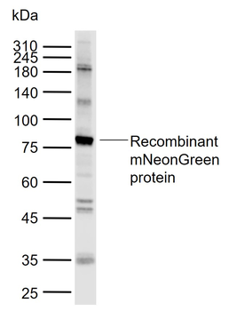
mNeonGreen Rabbit Polyclonal Antibody orb1291572.
Highly Specific Polyclonal
mNeonGreen Rabbit Polyclonal Antibody — orb1291572
orb1291572 is a robust rabbit polyclonal antibody explicitly raised against mNeonGreen. Because it binds multiple epitopes along the mNeonGreen structure, it offers exceptionally high sensitivity for detecting low-abundance fusions. It is ideal for Western Blot validation of expression constructs and endogenous CRISPR knock-ins where direct fluorescence is lost due to sample denaturation.
Host: Rabbit
Clonality: Polyclonal
Target: mNeonGreen
Reactivity: Branchiostoma lanceolatum / Engineered variants
Conjugation: Unconjugated
Supporting mNeonGreen Antibodies
Depending on your multiplexing constraints or need for high lot-to-lot consistency, Biorbyt also provides a highly specific mouse monoclonal option for precise assays.
SKU | Product | Applications |
|---|---|---|
mNeonGreen Rabbit Polyclonal Antibody Rabbit Polyclonal IgG | WB, ELISA, ICC/IF | |
mNeonGreen Mouse Monoclonal Antibody Mouse Monoclonal IgG | WB, IHC, ELISA |
6. Detecting and Validating mNeonGreen Constructs
Direct Fluorescence
mNeonGreen is routinely imaged using standard GFP or FITC filter sets. However, for maximum photon efficiency, utilize excitation near 506 nm and emission capture near 517 nm to take full advantage of its unique spectral profile.
Antibody Detection
When fluorescence cannot be preserved (e.g., in strict Western Blots or harsh IHC protocols), utilize Biorbyt's targeted orb1291572 (Rabbit) or orb2837255 (Mouse) antibodies to confirm correct molecular weight, expression, and fusion integrity.
7. Frequently Asked Questions
Is mNeonGreen a derivative of Aequorea victoria (jellyfish) GFP?
No. mNeonGreen is derived from a tetrameric yellow fluorescent protein found in the lancelet (Branchiostoma lanceolatum). Because of this, it shares only ~20% sequence homology with standard GFP.
Do I need special microscope filters to image mNeonGreen?
While optimized narrow-band filters centered around 506ex/517em will yield the absolute best signal-to-noise ratio, standard GFP or FITC filter cubes will successfully image mNeonGreen in most routine microscopy setups.
Can I use my standard anti-GFP antibody to detect mNeonGreen on a Western Blot?
Generally, no. Due to the lack of sequence identity, most pan-GFP antibodies will not bind mNeonGreen. You must use an antibody specifically raised against mNeonGreen, such as orb1291572 or orb2837255.
8. Scientific References
- Shaner NC, et al. A bright monomeric green fluorescent protein derived from Branchiostoma lanceolatum. Nature Methods. 2013;10(5):407-409. doi:10.1038/nmeth.2413
- Feng S, et al. Improved Split Fluorescent Proteins for Endogenous Protein Labeling. Nature Communications. 2017;8:370. doi:10.1038/s41467-017-00494-8
- Hostettler L, et al. The Bright Fluorescent Protein mNeonGreen Facilitates Protein Expression Analysis In Vivo. G3 (Bethesda). 2017;7(2):607-615. doi:10.1534/g3.116.038133
- Tanida-Miyake E, et al. Optimization of mNeonGreen for Homo sapiens increases its fluorescent intensity in mammalian cells. PLOS One. 2018;13(1):e0191108. doi:10.1371/journal.pone.0191108