You have no items in your shopping cart.
Featured
Description
Research Area
Signal Transduction
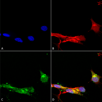
Images & Validation
−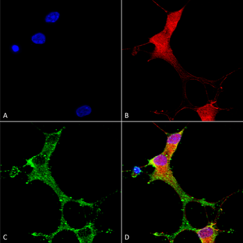
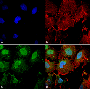
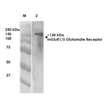
Item 1 of 3
| Tested Applications | ICC, IF, IHC, IP, WB |
|---|---|
| Dilution Range | WB (1:1000), IHC (1:1000), ICC/IF (1:1000) |
| Reactivity | Human, Mouse, Rat |
| Application Notes |
Key Properties
−| Host | Mouse |
|---|---|
| Clonality | Monoclonal |
| Isotype | IgG2A |
| Clone No. | N75/3 (Formerly sold as S75-3) |
| Immunogen | Fusion protein amino acids 824-1203 (cytoplasmic C-terminus) of rat mGluR5b |
| Target | mGluR5 |
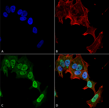
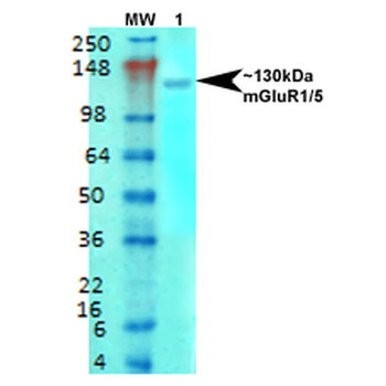
| Molecular Weight | 130kDa |
| Purification | Protein G Purified |
| Conjugation | FITC |
Storage & Handling
−| Storage | Conjugated antibodies should be stored according to the product label |
|---|---|
| Buffer/Preservatives | 640.91mM DMSO, 136.36 mM Ethanolamine, 126.89 mM chlorides, 9.09mM phosphates, 9.09mM NaHCO3 |
| Concentration | 1 mg/ml |
| Expiration Date | 12 months from date of receipt. |
| Disclaimer | For research use only |
Alternative Names
−mGluR5, mGlu5, GRM5, GPRC1E, Metabotropic glutamate receptor 5, Metabotropic glutamate receptor 5 variant F, Metabotropic glutamate receptor 5 variant G, Metabotropic glutamate receptor 5 variant H, Glutamate receptor metabotropic 5, mGluR5a, mGluR5b, mGluR, Glutamate receptor metabotropic 1, mGlu1, mGluR1, GRM1, GRM 1, GRM1A, GRM1 alpha, GRM1_HUMAN, GPRC1A, Metabotropic glutamate receptor 1, mGluR1 alpha
Similar Products
−- Item 1 of 3

MGluR5 Rabbit Polyclonal Antibody (FITC) [orb15702]
IF
Canine, Gallus, Human, Mouse, Sheep
Rat
Rabbit
Polyclonal
FITC
100 μlMetabotropic Glutamate Receptor 5/GRM5 Rabbit Polyclonal Antibody (FITC) [orb2589856]
Human, Rat
Rabbit
Polyclonal
FITC
100 μgMetabotropic Glutamate Receptor 5/GRM5 Rabbit Polyclonal Antibody (FITC) [orb2608546]
Human, Mouse, Rat
Rabbit
Polyclonal
FITC
100 μg
Quality Guarantee
Explore bioreagents carefree to elevate your research. All our products are rigorously tested for performance. If a product does not perform as described on its datasheet, our scientific support team will provide expert troubleshooting, a prompt replacement, or a refund. For full details, please see our Terms & Conditions and Buying Guide. Contact us at [email protected].
Quick Database Links
UniProt Details
− No UniProt data available
NCBI Gene Details
− No NCBI Gene data available
NCBI Reference Sequences
−Associated Accession Numbers
Curated reference sequences for the gene transcript and protein product| Protein | NP_058708.1 |
|---|
Protocol Information
WB
Western Blot (IB, immunoblot)
IHC
Immunohistochemistry
IF
Immunofluorescence
ICC
Immunocytochemistry
IP
Immunoprecipitation
Available Sizes
Select a size below
Choose Conjugation or Carrier Free Version
Free Secondary Antibody (20 ul)0/0
Please add an antibody product to your cart first.