You have no items in your shopping cart.
Description
Research Area
Immunology & Inflammation
Images & Validation
−

Item 1 of 2
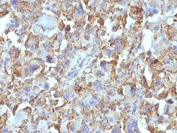
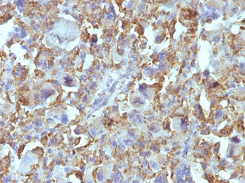
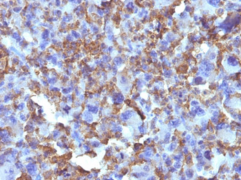
| Tested Applications | IHC-P |
|---|---|
| Dilution Range | Immunohistochemistry (FFPE): 0.5-1ug/ml for 30 min at RT |
| Reactivity | Human, Rat |
| Application Notes |
Key Properties
−| Antibody Type | Primary Antibody |
|---|---|
| Host | Mouse |
| Clonality | Monoclonal |
| Isotype | Mouse IgG1, kappa |
| Clone No. | D11 |
| Immunogen | A membrane preparation from human hepatocytes was used as the immunogen for the Macrophage / Histiocytoma Marker antibody. |
| Purification | Protein G affinity chromatography |
| Conjugation | Unconjugated |
Storage & Handling
−| Storage | Maintain refrigerated at 2-8°C for up to 2 weeks. For long term storage store at -20°C in small aliquots to prevent freeze-thaw cycles. |
|---|---|
| Buffer/Preservatives | 0.2 mg/ml in 1X PBS with 0.1 mg/ml rAlbumin and 0.05% sodium azide |
| Expiration Date | 12 months from date of receipt. |
| Disclaimer | For research use only |
Similar Products
−- Item 1 of 2
Macrophage & Histiocytoma Marker Antibody [orb389052]
IHC
Human
Mouse
Monoclonal
Unconjugated
20 μg, 100 μg, 100 μg (without BSA and Azide) - Item 1 of 2
Macrophage / Histiocytoma Marker Antibody [D11] [orb1252394]
IHC-P
Human
Mouse
Monoclonal
Unconjugated
100 μg - Item 1 of 2
Macrophage / Histiocytoma Marker Antibody [orb2639672]
IHC-P
Human, Rat
Mouse
Monoclonal
Unconjugated
7 ml - Item 1 of 2
Macrophage / Histiocytoma Marker Antibody [orb2639673]
IHC-P
Human, Rat
Mouse
Monoclonal
Unconjugated
100 μg - Item 1 of 2
Macrophage & Histiocytoma Marker Antibody - With BSA and Azide [orb1461520]
IHC-P
Human
Mouse
Monoclonal
Unconjugated
20 μg, 100 μg


![Macrophage / Histiocytoma Marker Antibody [D11]](/images/pub/media/catalog/product/NewWebsite/15/orb1252394_1.jpg)
![Macrophage / Histiocytoma Marker Antibody [D11]](/images/pub/media/catalog/product/NewWebsite/15/orb1252394_2.jpg)


Quality Guarantee
Explore bioreagents carefree to elevate your research. All our products are rigorously tested for performance. If a product does not perform as described on its datasheet, our scientific support team will provide expert troubleshooting, a prompt replacement, or a refund. For full details, please see our Terms & Conditions and Buying Guide. Contact us at [email protected].
Available Sizes
Select a size below
Choose Conjugation or Carrier Free Version
Free Secondary Antibody (20 ul)0/0
Please add an antibody product to your cart first.