You have no items in your shopping cart.
Description
Images & Validation
−Item 1 of 6
| Tested Applications | FACS, IF, IHC-P, WB |
|---|---|
| Dilution Range | Western blot: 1-2ug/ml,Flow cytometry: 1-2ug/million cells,Immunofluorescence: 1-2ug/ml,Immunohistochemistry (FFPE): 1-2ug/ml for 30 min at RT |
| Reactivity | Human, Rat |
| Application Notes |
Key Properties
−| Antibody Type | Primary Antibody |
|---|---|
| Host | Mouse |
| Clonality | Monoclonal |
| Isotype | Mouse IgG1, kappa |
| Clone No. | H1 |
| Immunogen | A cytoskeleton preparation containing Cytokeratin 8 was used as the immunogen for this antibody. |
| Purification | Protein G affinity chromatography |
| Conjugation | Unconjugated |
Storage & Handling
−| Storage | Maintain refrigerated at 2-8°C for up to 2 weeks. For long term storage store at -20°C in small aliquots to prevent freeze-thaw cycles. |
|---|---|
| Buffer/Preservatives | 0.2 mg/ml in 1X PBS with 0.1 mg/ml rAlbumin and 0.05% sodium azide |
| Expiration Date | 12 months from date of receipt. |
| Disclaimer | For research use only |
Similar Products
−- Item 1 of 22
Cytokeratin 8 Mouse Monoclonal Antibody [orb500822]
FC, IF, IHC-Fr, IHC-P
Mouse, Rat
Human, Mouse, Rat
Mouse
Monoclonal
Unconjugated
50 μl, 100 μl, 200 μl, 200 μg - Item 1 of 16
Cytokeratin 8/KRT8 Rabbit Polyclonal Antibody [orb19070]
IHC, WB
Human, Mouse, Rat
Rabbit
Polyclonal
Unconjugated
100 μg - Item 1 of 10
Cytokeratin 18 Antibody [orb2638771]
FACS, IF, IHC-P, WB
Human, Rat
Mouse
Monoclonal
Unconjugated
100 μg - Item 1 of 10
Cytokeratin 18 Antibody [orb749637]
FACS, IF, IHC-P, WB
Human, Rat
Mouse
Monoclonal
Unconjugated
100 μg, 20 μg - Item 1 of 7
Cytokeratin 8 Rabbit Polyclonal Antibody [orb10413]
ELISA, IF, IHC-Fr, IHC-P
Gallus, Mouse, Rat
Human
Rabbit
Polyclonal
Unconjugated
100 μl, 50 μl, 200 μl


Quality Guarantee
Explore bioreagents carefree to elevate your research. All our products are rigorously tested for performance. If a product does not perform as described on its datasheet, our scientific support team will provide expert troubleshooting, a prompt replacement, or a refund. For full details, please see our Terms & Conditions and Buying Guide. Contact us at [email protected].
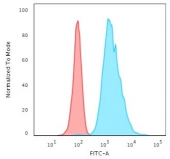
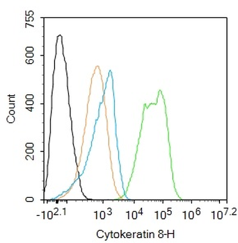
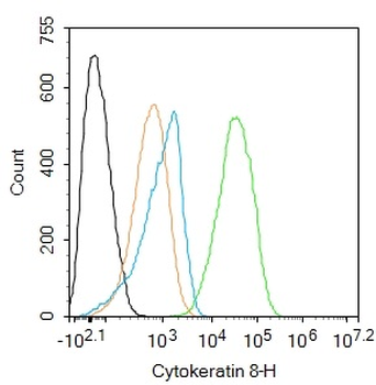
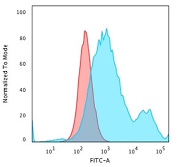
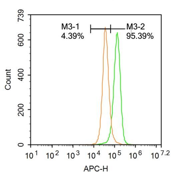
Flow cytometry testing of permeabilized human HeLa cells with Cytokeratin 8 antibody (clone H1); Red = isotype control, Blue = Cytokeratin 8 antibody.
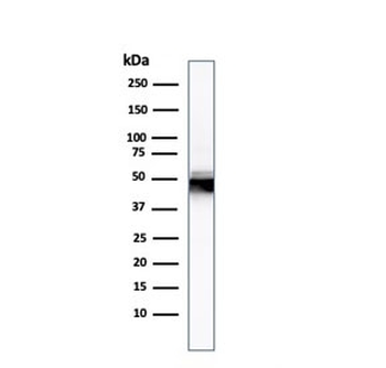
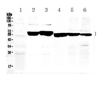
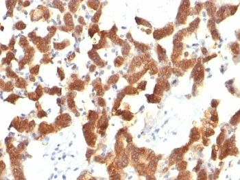
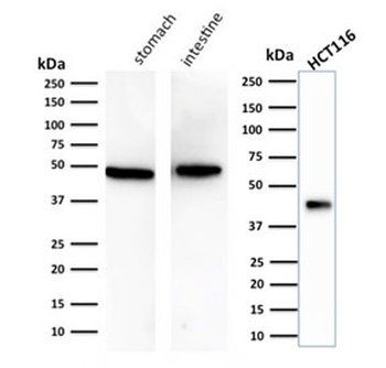
Western blot testing of human HCT-116 lysate with Cytokeratin 8 antibody.
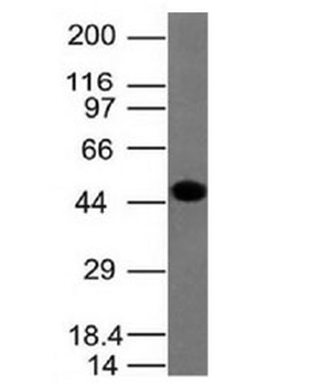
Western blot testing of A431 lysate with Cytokeratin 8 antibody
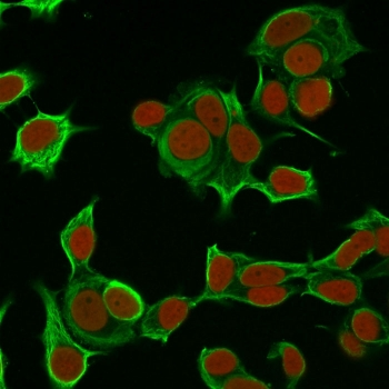
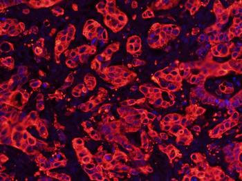
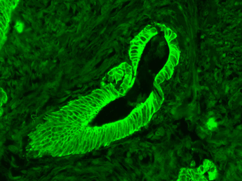
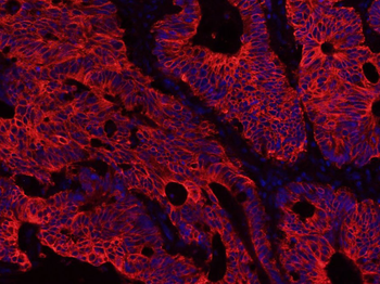
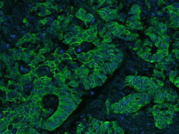
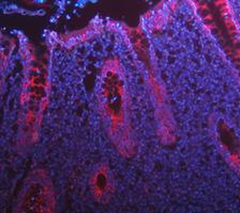
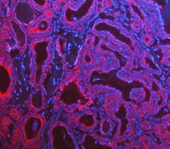
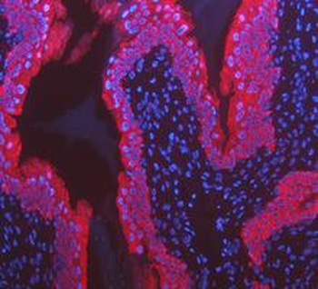
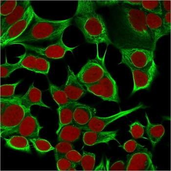
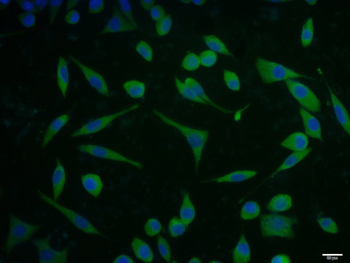
Immunofluorescent staining of permeabilized human MCF7 cells with Cytokeratin 8 antibody (clone H1, green) and Reddot nuclear stain (red).
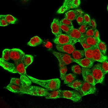
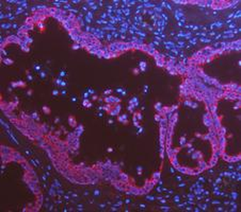
Immunofluorescent staining of permeabilized human HCT-116 cells with Cytokeratin 8 antibody (clone H1, green) and Reddot nuclear stain (red).
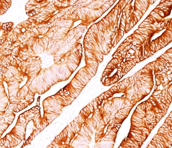
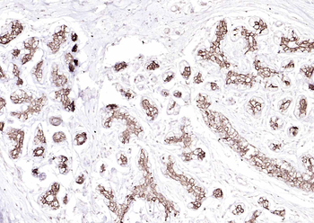
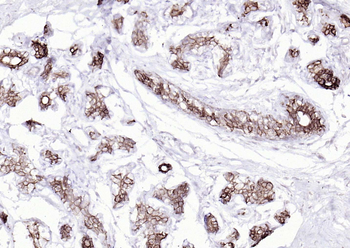
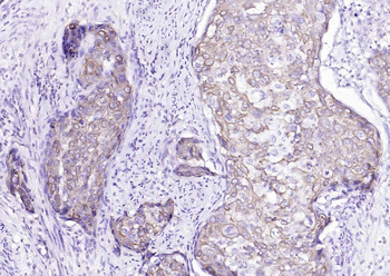
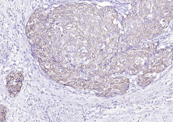
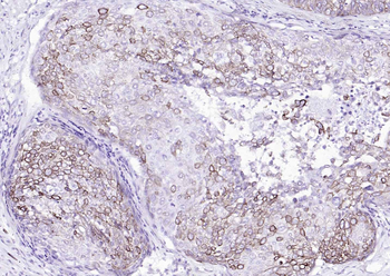
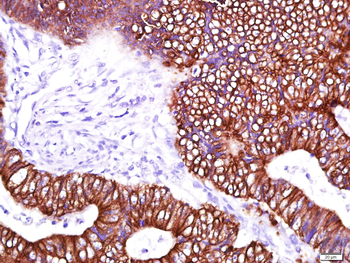
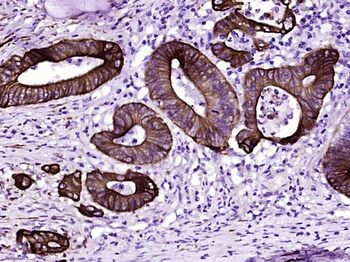
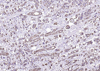
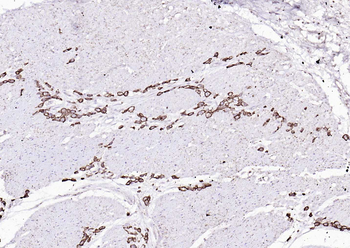
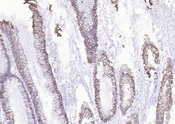
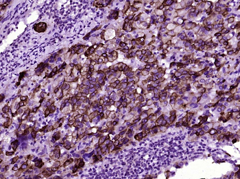
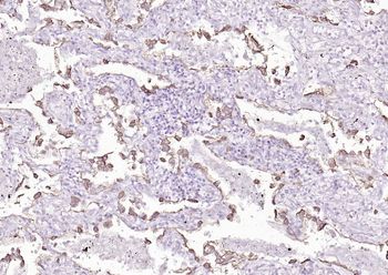
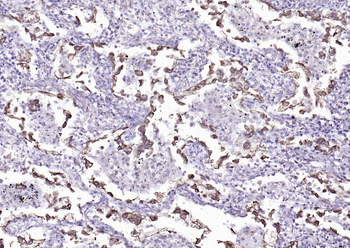
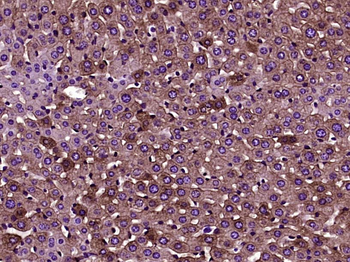
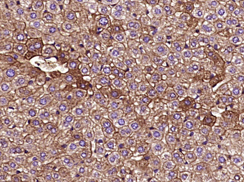
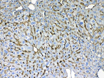
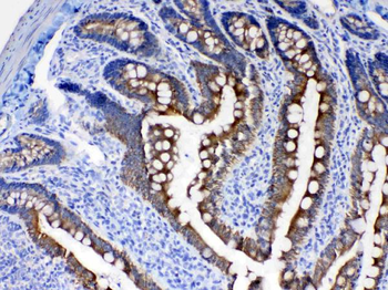
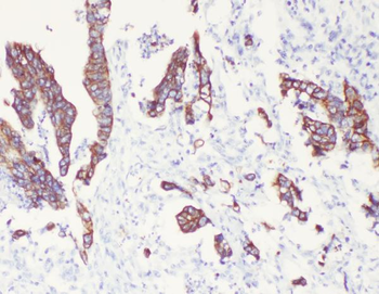
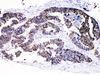
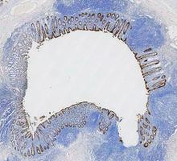
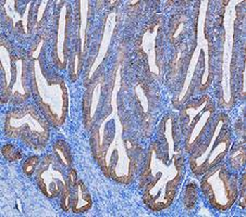
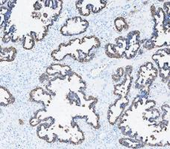
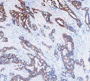
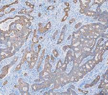
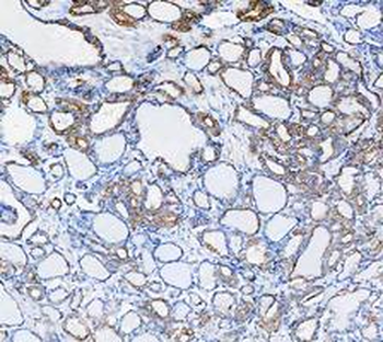
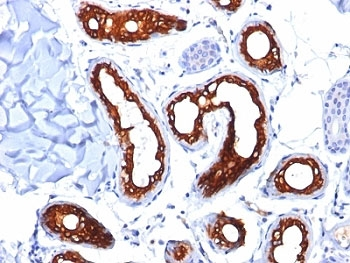
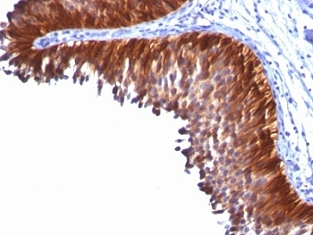
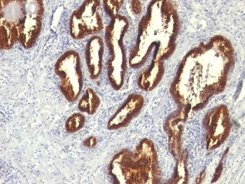
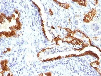
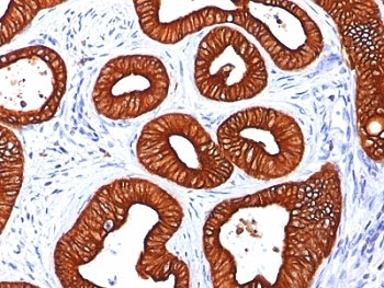
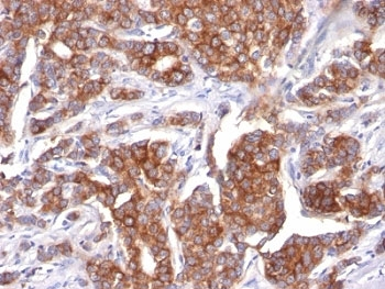
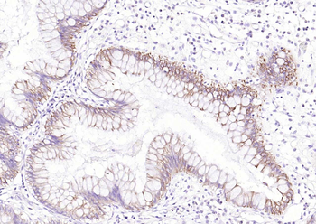
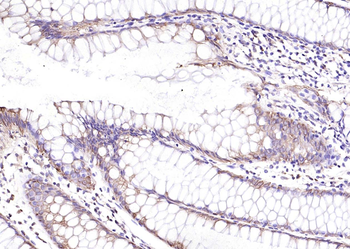
IHC testing of human colon carcinoma stained with Cytokeratin 8 antibody
Documents Download
Datasheet
Product Information
Request a Document
Protocol Information
WB
Western Blot (IB, immunoblot)
IHC-P
Immunohistochemistry Paraffin
FACS
Fluorescence-Activated Cell Sorting (FC, Flow cytometry)
IF
Immunofluorescence
Cytokeratin 8 Antibody (orb248434)

- 0.0
Based on 0 reviews
Participating in our Biorbyt product reviews program enables you to support fellow scientists by sharing your firsthand experience with our products.
Login to Submit a ReviewAvailable Sizes
Select a size below
Choose Conjugation or Carrier Free Version
Free Secondary Antibody (20 ul)0/0
Please add an antibody product to your cart first.