You have no items in your shopping cart.
Description
Research Area
Cell Biology, Disease Biomarkers, Protein Biochemistry, Signal Transduction
Images & Validation
−Item 1 of 3
| Tested Applications | ICC, IF, IHC, WB |
|---|---|
| Dilution Range | WB (1:1000), ICC/IF (1:100) |
| Reactivity | Human, Mouse, Rat |
| Application Notes |
Key Properties
−| Host | Rabbit |
|---|---|
| Clonality | Polyclonal |
| Immunogen | KDEL containing peptide immunogen |
| Target | KDEL |
| Purification | Protein A Purified |
| Conjugation | FITC |
Storage & Handling
−| Storage | Conjugated antibodies should be stored according to the product label |
|---|---|
| Buffer/Preservatives | 640.91mM DMSO, 136.36 mM Ethanolamine, 126.89 mM chlorides, 9.09mM phosphates, 9.09mM NaHCO3 |
| Concentration | 1 mg/ml |
| Expiration Date | 12 months from date of receipt. |
| Disclaimer | For research use only |
Alternative Names
−KDEL, K-D-E-L, Lys-Asp-Glu-Leu, Lys-Asp-Glu-Leu (KDEL), Lysine-aspartic acid-glutamate-leucine
Similar Products
−- Item 1 of 4
Calreticulin Antibody (FITC) [orb151514]
FC, ICC, IF, IHC, IP, WB
Bovine, Canine, Gallus, Guinea pig, Hamster, Human, Monkey, Mouse, Porcine, Rabbit, Rat, Sheep
Rabbit
Polyclonal
FITC
200 μl - Item 1 of 3
PDI Antibody (FITC) [orb151395]
ICC, IF, IHC, IP, WB
Bovine, Canine, Frog, Guinea pig, Hamster, Human, Invertebrate, Mouse, Mussel, Porcine, Rat, Sheep
Rabbit
Polyclonal
FITC
100 μl - Item 1 of 3
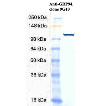
GRP94 Antibody (FITC) [orb151208]
ICC, IF, IHC, IP, WB
Bovine, Human, Mouse, Rat
Rabbit
Polyclonal
FITC
100 μg - Item 1 of 3
GRP94 Antibody (FITC) [orb146770]
FC, ICC, IF, IP, WB
Bovine, Canine, Equine, Frog, Gallus, Guinea pig, Hamster, Human, Monkey, Mouse, Porcine, Rabbit, Rat, Sheep
Rat
Monoclonal
FITC
200 μg - Item 1 of 2

Quality Guarantee
Explore bioreagents carefree to elevate your research. All our products are rigorously tested for performance. If a product does not perform as described on its datasheet, our scientific support team will provide expert troubleshooting, a prompt replacement, or a refund. For full details, please see our Terms & Conditions and Buying Guide. Contact us at [email protected].
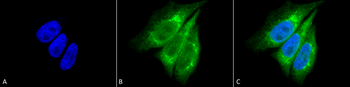
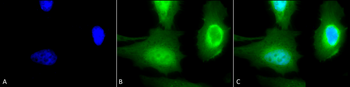
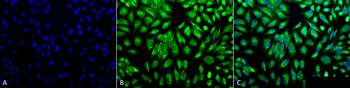
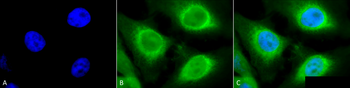
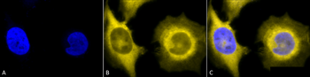
Immunocytochemistry/Immunofluorescence analysis using Rabbit Anti-KDEL Polyclonal Antibody. Tissue: Heat Shocked Cervical cancer cell line (HeLa). Species: Human. Fixation: 2% Formaldehyde for 20 min at RT. Primary Antibody: Rabbit Anti-KDEL Polyclonal Antibody at 1:100 for 12 hours at 4°C. Secondary Antibody: FITC Goat Anti-Rabbit (green) at 1:200 for 2 hours at RT. Counterstain: DAPI (blue) nuclear stain at 1:40000 for 2 hours at RT. Localization: Endoplasmic reticulum. Magnification: 100x. (A) DAPI (blue) nuclear stain. (B) Anti-KDEL Antibody. (C) Composite. Heat Shocked at 42°C for 30 min.

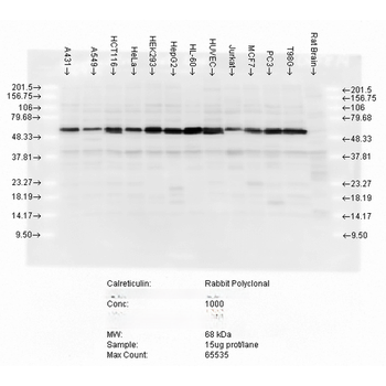
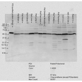
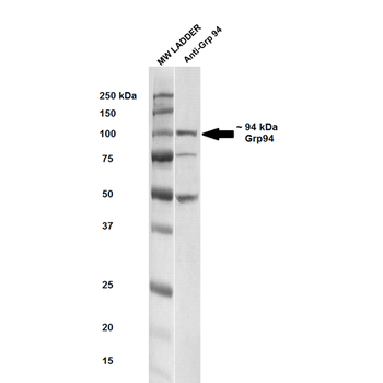
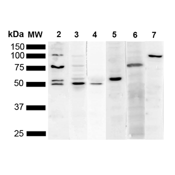
Western blot analysis of Human Cell line lysates showing detection of KDEL protein using Rabbit Anti-KDEL Polyclonal Antibody. Primary Antibody: Rabbit Anti-KDEL Polyclonal Antibody at 1:1000, 1:500.
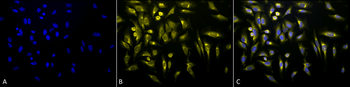
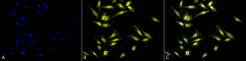
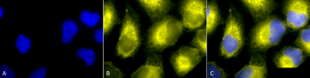
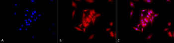
Immunocytochemistry/Immunofluorescence analysis using Rabbit Anti-KDEL Polyclonal Antibody. Tissue: Heat Shocked Cervical cancer cell line (HeLa). Species: Human. Fixation: 2% Formaldehyde for 20 min at RT. Primary Antibody: Rabbit Anti-KDEL Polyclonal Antibody at 1:100 for 12 hours at 4°C. Secondary Antibody: R-PE Goat Anti-Rabbit (yellow) at 1:200 for 2 hours at RT. Counterstain: DAPI (blue) nuclear stain at 1:40000 for 2 hours at RT. Localization: Endoplasmic reticulum. Magnification: 20x. (A) DAPI (blue) nuclear stain. (B) Anti-KDEL Antibody. (C) Composite. Heat Shocked at 42°C for 30 min.
Quick Database Links
Gene Symbol
KDEL
Documents Download
Datasheet
Product Information
Request a Document
Protocol Information
WB
Western Blot (IB, immunoblot)
IHC
Immunohistochemistry
IF
Immunofluorescence
ICC
Immunocytochemistry
KDEL Antibody (FITC) (orb151344)

- 0.0
Based on 0 reviews
Participating in our Biorbyt product reviews program enables you to support fellow scientists by sharing your firsthand experience with our products.
Login to Submit a ReviewAvailable Sizes
Select a size below
Free Secondary Antibody (20 ul)0/0
Please add an antibody product to your cart first.