You have no items in your shopping cart.
Description
Research Area
Protein Biochemistry
Images & Validation
−Item 1 of 1
| Application Notes |
|---|
Key Properties
−| Source | Human |
|---|---|
| Biological Origin | Human |
| Biological Activity | 4.5 |
| Isotype | Albumin |
| Conjugation | Texas Red |
| Purity | This product was prepared from normal serum by a multi-step process including selective precipitation and extensive dialysis against the buffer stated above. Assay by immunoelectrophoresis resulted in a single precipitin arc against anti-Human Serum. |
Storage & Handling
−| Storage | Store vial at 4° C prior to restoration. For extended storage aliquot contents and freeze at -20° C or below. Avoid cycles of freezing and thawing. Centrifuge product if not completely clear after standing at room temperature. This product is stable for several weeks at 4° C as an undiluted liquid. Dilute only prior to immediate use. |
|---|---|
| Form/Appearance | Lyophilized |
| Buffer/Preservatives | Preservative: 0.01% (w/v) Sodium Azide. Stabilizer: 10 mg/ml Polyethylene Glycol (PEG-8000); Buffer: 0.02 M Potassium Phosphate, 0.15 M Sodium Chloride, pH 7.2 |
| Concentration | 1.0 mg/ml |
| Expiration Date | 6 months from date of receipt. |
| Disclaimer | For research use only |
Alternative Names
−Human Albumin Texas Red conjugation
Quality Guarantee
Explore bioreagents carefree to elevate your research. All our products are rigorously tested for performance. If a product does not perform as described on its datasheet, our scientific support team will provide expert troubleshooting, a prompt replacement, or a refund. For full details, please see our Terms & Conditions and Buying Guide. Contact us at [email protected].
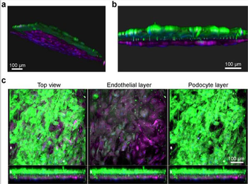
Fluorescence microscopy images of the human kidney Glomerulus Chip established from iPS cell-derived podocytes and primary glomerular endothelial cells. (a) Side and (b) cross-sectional view of 3D reconstructed confocal images of the human Glomerulus Chip showing the iPS cell-derived podocytes and endothelial cells in their respective layers after differentiation and co-culture on opposing sides of the flexible ECM-coated PDMS membrane. (c) Additional immunofluorescence confocal images showing a top view of both cell layers (left), the endothelial cell layer only (middle), and the human iPS cell-derived podocyte layer (right). Scale bars, 100 µm. Human albumin conjugated to Texas Red.
Quick Database Links
UniProt
RefSeq:AAA98797.1
UniProt Details
− No UniProt data available
NCBI Reference Sequences
−Associated Accession Numbers
Curated reference sequences for the gene transcript and protein product| RefSeq | AAA98797.1 |
|---|
Documents Download
Datasheet
Product Information
Request a Document
Protocol Information
Protein Handling and Storage Guide
Protein Handling Guide
Human Albumin protein (Texas Red) (orb346270)

- 0.0
Based on 0 reviews
Participating in our Biorbyt product reviews program enables you to support fellow scientists by sharing your firsthand experience with our products.
Login to Submit a ReviewAvailable Sizes
Select a size below