You have no items in your shopping cart.
Featured
Description
Research Area
Epigenetics & Chromatin
Images & Validation
−Item 1 of 5
| Tested Applications | ELISA, Enzyme Assay, ICC, IF, IHC, IP, WB |
|---|---|
| Dilution Range | WB (1:1000), IHC-P (1:2000), ICC/IF (1:100) |
| Reactivity | Bovine, Guinea pig, Hamster, Human, Monkey, Mouse, Rabbit, Rat |
| Application Notes |
Key Properties
−| Host | Rat |
|---|---|
| Clonality | Monoclonal |
| Isotype | IgG1 |
| Clone No. | 4B4 |
| Immunogen | Purified recombinant mouse HSF1 protein, epitope mapping to amino acids 425-439 |
| Target | HSF1 |
| Molecular Weight | 95kDa |
| Purification | Protein G Purified |
| Conjugation | Unconjugated |
Storage & Handling
−| Storage | Maintain refrigerated at 2-8°C for up to 2 weeks. For long term storage store at -20°C in small aliquots to prevent freeze-thaw cycles. |
|---|---|
| Buffer/Preservatives | PBS pH 7.4, 50% glycerol, 0.1% sodium azide. Storage buffer changes when conjugated. |
| Concentration | 1 mg/ml |
| Expiration Date | 12 months from date of receipt. |
| Disclaimer | For research use only |
Alternative Names
−HSF1, Heat shock factor protein 1, Heat shock transcription factor 1, HSTF1, HSF 1
Similar Products
−- Item 1 of 16
HSF1 Rabbit Polyclonal Antibody [orb5463]
ICC, IF, IHC-Fr, IHC-P
Bovine, Canine, Porcine, Rabbit
Human, Mouse, Rat
Rabbit
Polyclonal
Unconjugated
50 μl, 100 μl, 200 μl - Item 1 of 9
Phospho-HSF1 (Ser326) Recombinant Rabbit Monoclonal Antibody [orb559454]
FC, IF, IHC-Fr, IHC-P, WB
Rat
Human
Rabbit
Recombinant
Unconjugated
50 μl, 100 μl, 200 μl, 200 μg, 25 μl - Item 1 of 9
- Item 1 of 6
- Item 1 of 5
Phospho-HSF1 (Ser303 + Ser307) Rabbit Polyclonal Antibody [orb184051]
IF, IHC-Fr, IHC-P, WB
Bovine, Canine, Rabbit
Human, Mouse, Rat
Rabbit
Polyclonal
Unconjugated
200 μl, 50 μl, 100 μl


Quality Guarantee
Explore bioreagents carefree to elevate your research. All our products are rigorously tested for performance. If a product does not perform as described on its datasheet, our scientific support team will provide expert troubleshooting, a prompt replacement, or a refund. For full details, please see our Terms & Conditions and Buying Guide. Contact us at [email protected].
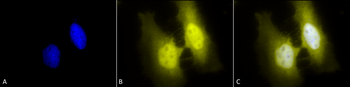
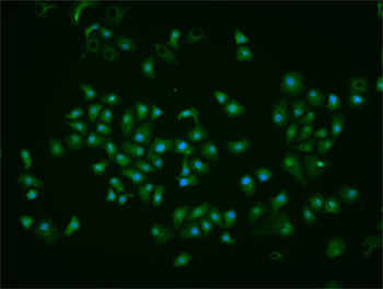
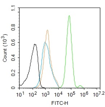
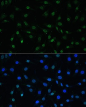
Immunocytochemistry/Immunofluorescence analysis using Rat Anti-HSF1 Monoclonal Antibody, Clone 4B4. Tissue: Heat Shocked cervical cancer cells (HeLa). Species: Human. Fixation: 2% Formaldehyde for 20 min at RT. Primary Antibody: Rat Anti-HSF1 Monoclonal Antibody at 1:100 for 12 hours at 4°C. Secondary Antibody: R-PE Goat Anti-Rat (yellow) at 1:200 for 2 hours at RT. Counterstain: DAPI (blue) nuclear stain at 1:40000 for 2 hours at RT. Localization: Cytoplasm. Localizes to the nucleus upon activation. Magnification: 100x. (A) DAPI (blue) nuclear stain. (B) Anti-HSF1 Antibody. (C) Composite. Heat Shocked at 42°C for 1h.
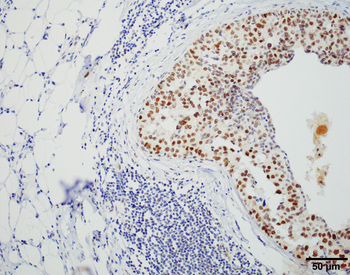
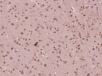
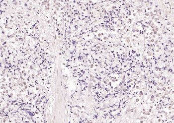
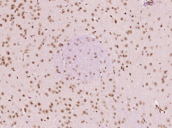
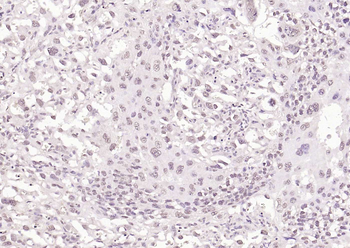
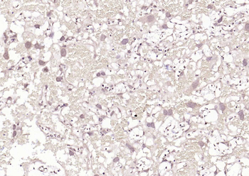
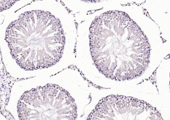
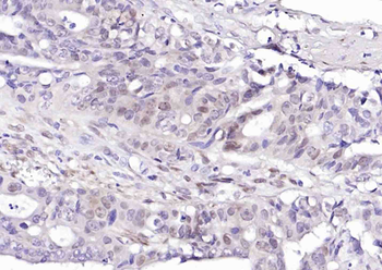
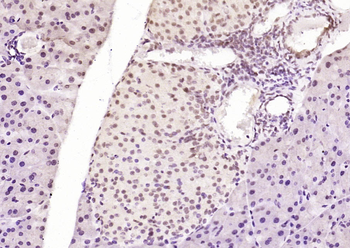
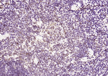
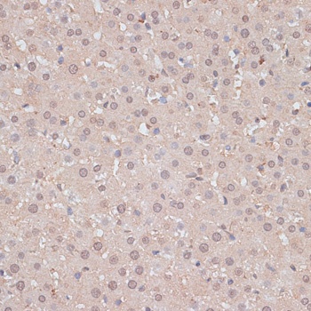
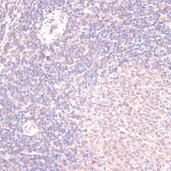
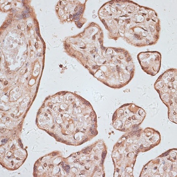
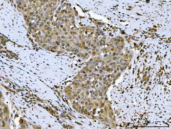
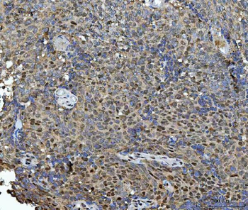
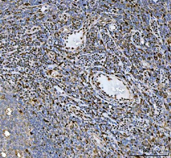
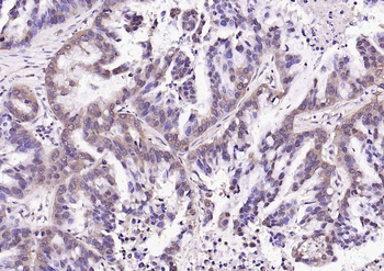
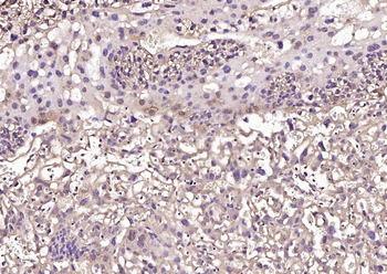
Immunohistochemistry analysis using Rat Anti-HSF1 Monoclonal Antibody, Clone 4B4. Tissue: Breast carcinoma. Species: Human. Fixation: 10% Formalin Solution for 20 hours at RT. Primary Antibody: Rat Anti-HSF1 Monoclonal Antibody at 1:2000 for 40 min. Secondary Antibody: Dako labeled Polymer HRP Anti-rat IgG, DAB Chromogen (brown) (Dako Envision+ System) for 30 min at RT. Counterstain: Mayer's Hematoxylin (purple/blue) nuclear stain for 1 minute at RT. Localization: Nuclear. Magnification: 100X.
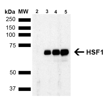
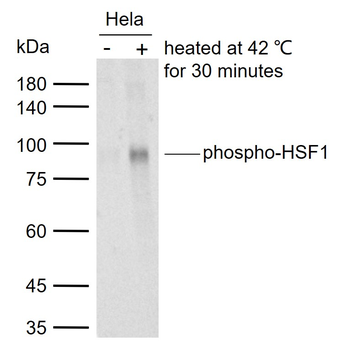
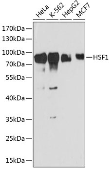
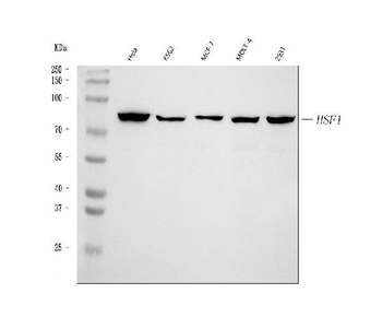
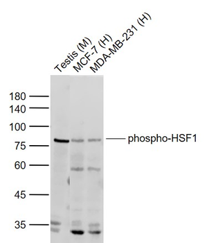
Western Blot analysis of Human Breast adenocarcinoma cell line (MCF7) showing detection of ~65 kDa HSF1 protein using Rat Anti-HSF1 Monoclonal Antibody, Clone 4B4. Lane 1: MW ladder. Lane 2: HSF1 null lysate prepared from mouse embryonic fibroblasts. Lane 3: MCF7 lysate (5 μg). Lane 4: MCF7 lysate (10 μg). Lane 5: MCF7 lysate (20 μg). Block: 1.5% BSA for 30 minutes at RT. Primary Antibody: Rat Anti-HSF1 Monoclonal Antibody at 1:1000 for 2 hours at RT. Secondary Antibody: Goat Anti-Rat IgG: HRP for 1 hour at RT. Predicted/Observed Size: ~65 kDa.
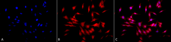
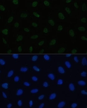
Immunocytochemistry/Immunofluorescence analysis using Rat Anti-HSF1 Monoclonal Antibody, Clone 4B4. Tissue: Heat Shocked cervical cancer cells (HeLa). Species: Human. Fixation: 2% Formaldehyde for 20 min at RT. Primary Antibody: Rat Anti-HSF1 Monoclonal Antibody at 1:100 for 12 hours at 4°C. Secondary Antibody: APC Goat Anti-Rat (red) at 1:200 for 2 hours at RT. Counterstain: DAPI (blue) nuclear stain at 1:40000 for 2 hours at RT. Localization: Cytoplasm. Localizes to the nucleus upon activation. Magnification: 20x. (A) DAPI (blue) nuclear stain. (B) Anti-HSF1 Antibody. (C) Composite. Heat Shocked at 42°C for 1h.
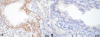
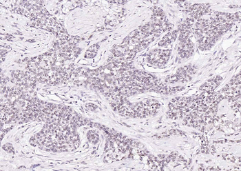
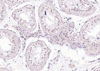
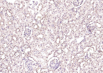
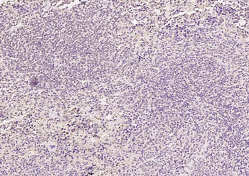
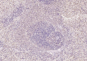
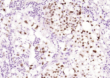
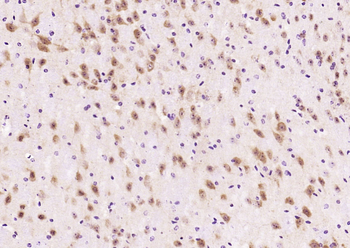
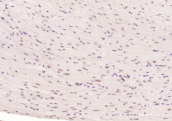
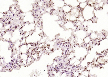
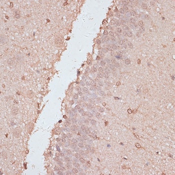
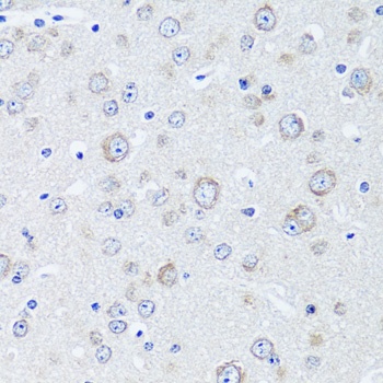
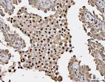
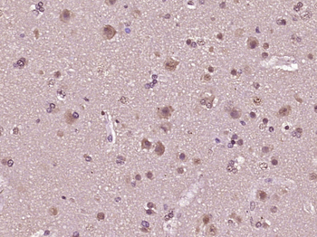
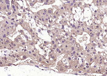
Immunohistochemistry analysis using Rat Anti-HSF1 Monoclonal Antibody, Clone 4B4. Tissue: Lung. Species: Mouse. Fixation: 10% Formalin Solution for 20 hours at RT. Primary Antibody: Rat Anti-HSF1 Monoclonal Antibody at 1:1000 for 40 min. Secondary Antibody: Dako labeled Polymer HRP Anti-rat IgG, DAB Chromogen (brown) (Dako Envision+ System) for 30 min at RT. Counterstain: Mayer's Hematoxylin (purple/blue) nuclear stain for 1 minute at RT. Localization: Nuclear. Magnification: 100X. (A) HSF Wildtype. (B) HSF null.
Quick Database Links
UniProt Details
− No UniProt data available
NCBI Gene Details
− No NCBI Gene data available
NCBI Reference Sequences
−Associated Accession Numbers
Curated reference sequences for the gene transcript and protein product| Protein | NP_032322.1 |
|---|
Documents Download
Datasheet
Product Information
Request a Document
Protocol Information
WB
Western Blot (IB, immunoblot)
IHC
Immunohistochemistry
IF
Immunofluorescence
ICC
Immunocytochemistry
ELISA
Enzyme-linked Immunosorbent Assay (EIA)
IP
Immunoprecipitation
HSF1 Antibody (orb151126)

- 0.0
Based on 0 reviews
Participating in our Biorbyt product reviews program enables you to support fellow scientists by sharing your firsthand experience with our products.
Login to Submit a ReviewAvailable Sizes
Select a size below
Choose Conjugation or Carrier Free Version
Free Secondary Antibody (20 ul)0/0
Please add an antibody product to your cart first.