You have no items in your shopping cart.
Featured
Description
Research Area
Cell Biology
Images & Validation
−Item 1 of 4
| Tested Applications | AM, ELISA, ICC, IF, IHC, IP, PBA, PLA, WB |
|---|---|
| Dilution Range | WB (1:1000), ICC/IF (1:100); optimal dilutions for assays should be determined by the user. |
| Reactivity | Human, Mouse, Rat |
| Application Notes |
Key Properties
−| Host | Mouse |
|---|---|
| Clonality | Monoclonal |
| Isotype | IgG2a Kappa |
| Clone No. | 1F2-H5 |
| Immunogen | Full length human HSC70 |
| Target | HSC70 (HSP73) |
| Molecular Weight | 73kDa |
| Purification | Protein G Purified |
| Conjugation | Unconjugated |
Storage & Handling
−| Storage | Maintain refrigerated at 2-8°C for up to 2 weeks. For long term storage store at -20°C in small aliquots to prevent freeze-thaw cycles. |
|---|---|
| Buffer/Preservatives | PBS pH 7.4, 50% glycerol, 0.09% sodium azide. Storage buffer changes when conjugated. |
| Concentration | 1 mg/ml |
| Expiration Date | 12 months from date of receipt. |
| Disclaimer | For research use only |
Alternative Names
−HSPA8, HSC70, HSP73, Heat shock cognate 71 kDa protein, Heat shock 70 kDa protein 8, HSP71, HSC71, HSC54, HSC73, HSPA10, LAP1, NIP71
Similar Products
−- Item 1 of 12
HSC70 Mouse Monoclonal Antibody [orb704174]
IF, IHC-Fr, IHC-P, WB
Mouse, Rat
Human, Mouse, Rat
Mouse
Monoclonal
Unconjugated
100 μl, 50 μl, 200 μl, 200 μg - Item 1 of 12
Hsc70 Mouse Monoclonal Antibody [orb623830]
FC, ICC, IF, IHC, WB
Human, Mouse, Rat
Mouse
Monoclonal
Unconjugated
100 μg - Item 1 of 12
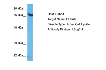
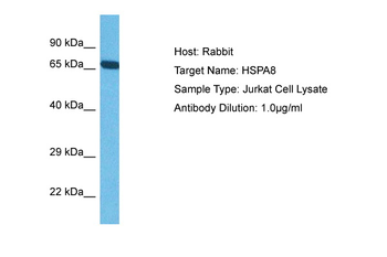
HSPA8 Rabbit Polyclonal Antibody [orb330554]
IHC, WB
Bovine, Goat, Porcine, Sheep, Zebrafish
Human, Mouse, Rat
Rabbit
Polyclonal
Unconjugated
100 μl - Item 1 of 9
- Item 1 of 7
HSPA8 Antibody [orb688870]
ELISA, FC, IHC, IP, WB
Human, Mouse, Rat
Mouse
Monoclonal
Unconjugated
50 μl, 100 μl

Quality Guarantee
Explore bioreagents carefree to elevate your research. All our products are rigorously tested for performance. If a product does not perform as described on its datasheet, our scientific support team will provide expert troubleshooting, a prompt replacement, or a refund. For full details, please see our Terms & Conditions and Buying Guide. Contact us at [email protected].

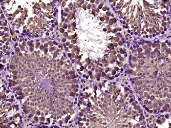
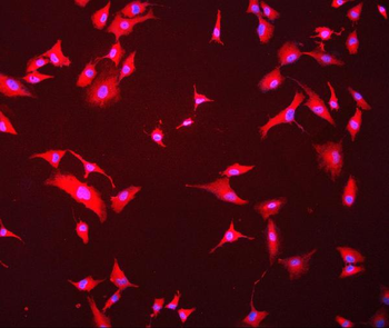
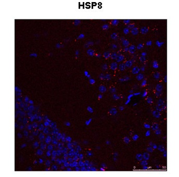
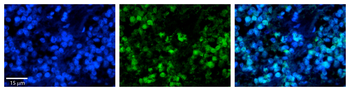
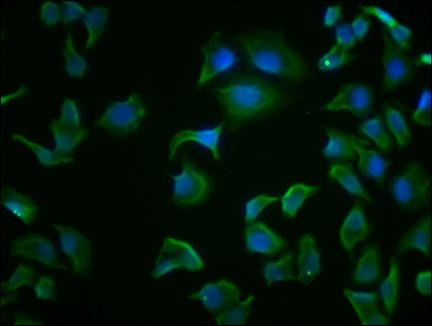
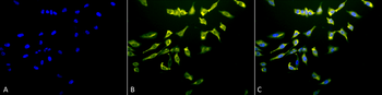
Immunocytochemistry/Immunofluorescence analysis using Mouse Anti-Hsc70 (Hsp73) Monoclonal Antibody, Clone 1F2-H5. Tissue: Heat Shocked cervical cancer cells (HeLa). Species: Human. Fixation: 2% Formaldehyde for 20 min at RT. Primary Antibody: Mouse Anti-Hsc70 (Hsp73) Monoclonal Antibody at 1:100 for 12 hours at 4°C. Secondary Antibody: FITC Goat Anti-Mouse (green) at 1:200 for 2 hours at RT. Counterstain: DAPI (blue) nuclear stain at 1:40000 for 2 hours at RT. Localization: Cytoplasm. Melanosome. Localizes to nucleus upon heat shock. Magnification: 100x. (A) DAPI (blue) nuclear stain. (B) Anti-Hsc70 (Hsp73) Antibody. (C) Composite. Heat Shocked at 42°C for 1h.
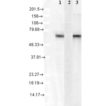
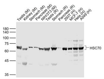
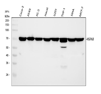
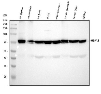
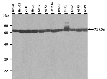
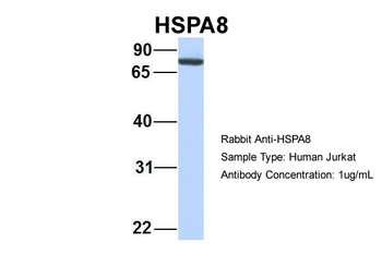
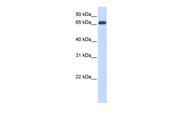
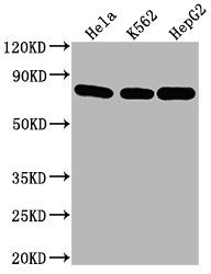
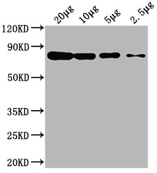
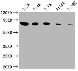
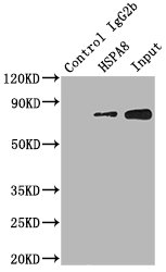
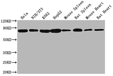
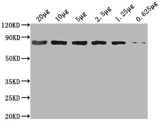
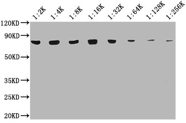
Western Blot analysis of Human Cell lysates showing detection of Hsc70 protein using Mouse Anti-Hsc70 Monoclonal Antibody, Clone 1F2-H5. Load: 15 μg. Block: 1.5% BSA for 30 minutes at RT. Primary Antibody: Mouse Anti-Hsc70 Monoclonal Antibody at 1:1000 for 2 hours at RT. Secondary Antibody: Sheep Anti-Mouse IgG: HRP for 1 hour at RT. 1: mix of 10 different human cell lines, 2: Hsp72 recombinant protein, and 3: Hsc70 (Hsp73) recombinant protein.
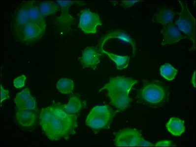
Immunocytochemistry/Immunofluorescence analysis using Mouse Anti-Hsc70 (Hsp73) Monoclonal Antibody, Clone 1F2-H5. Tissue: Heat Shocked cervical cancer cells (HeLa). Species: Human. Fixation: 2% Formaldehyde for 20 min at RT. Primary Antibody: Mouse Anti-Hsc70 (Hsp73) Monoclonal Antibody at 1:100 for 12 hours at 4°C. Secondary Antibody: R-PE Goat Anti-Mouse (yellow) at 1:200 for 2 hours at RT. Counterstain: DAPI (blue) nuclear stain at 1:40000 for 2 hours at RT. Localization: Cytoplasm. Melanosome. Localizes to nucleus upon heat shock. Magnification: 20x. (A) DAPI (blue) nuclear stain. (B) Anti-Hsc70 (Hsp73) Antibody. (C) Composite. Heat Shocked at 42°C for 1h.
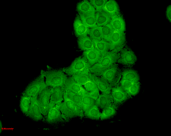
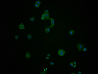
Immunocytochemistry/Immunofluorescence analysis using Mouse Anti-Hsc70 Monoclonal Antibody, Clone 1F2-H5. Tissue: HaCaT cells. Species: Human. Fixation: Cold 100% methanol for 10 minutes at -20°C. Primary Antibody: Mouse Anti-Hsc70 Monoclonal Antibody at 1:100 for 1 hour at RT. Secondary Antibody: FITC Goat Anti-Mouse (green) at 1:50 for 1 hour at RT. Localization: Bright cytoplasmic staining, duller nuclear staining.
Quick Database Links
UniProt Details
− No UniProt data available
NCBI Gene Details
− No NCBI Gene data available
NCBI Reference Sequences
−Associated Accession Numbers
Curated reference sequences for the gene transcript and protein product| Protein | NP_006588.1 |
|---|
Documents Download
Datasheet
Product Information
Request a Document
Protocol Information
WB
Western Blot (IB, immunoblot)
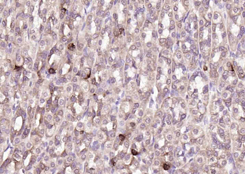
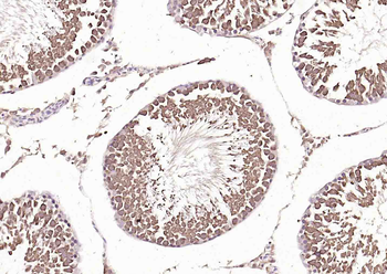
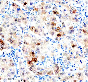
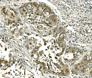
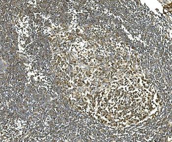
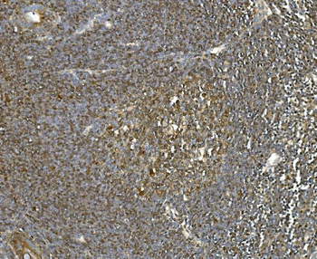
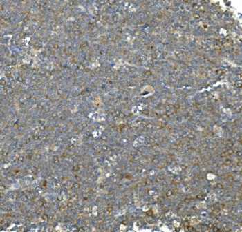
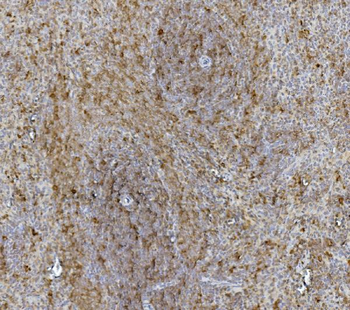
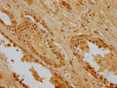
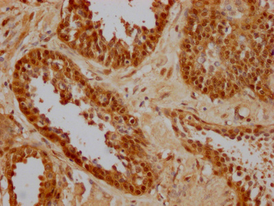
IHC
Immunohistochemistry
IF
Immunofluorescence
ICC
Immunocytochemistry
ELISA
Enzyme-linked Immunosorbent Assay (EIA)
IP
Immunoprecipitation
HSC70 (HSP73) Antibody (orb1822478)

- 0.0
Based on 0 reviews
Participating in our Biorbyt product reviews program enables you to support fellow scientists by sharing your firsthand experience with our products.
Login to Submit a ReviewAvailable Sizes
Select a size below
Free Secondary Antibody (20 ul)0/0
Please add an antibody product to your cart first.