You have no items in your shopping cart.
Description
Research Area
Pharmacology & Drug Discovery
Images & Validation
−Item 1 of 5
| Tested Applications | ELISA, FACS, FC, ICC, IF, WB |
|---|---|
| Dilution Range | WB (1:1000); ICC/IF (1:50); ELISA (1:1000); FACS (1:50); FCM (1:50) |
| Reactivity | All |
| Application Notes |
Key Properties
−| Host | Mouse |
|---|---|
| Clonality | Monoclonal |
| Isotype | IgG1 |
| Clone No. | 500000000 |
| Immunogen | Synthetic Hexanoyl modified Keyhole Limpet Hemocyanin (KLH). |
| Target | Hexanoyl-Lysine adduct |
| Purification | Protein G Purified |
| Conjugation | Biotin |
Storage & Handling
−| Storage | Conjugated antibodies should be stored according to the product label |
|---|---|
| Buffer/Preservatives | 136.36mM Ethanolamine, 133.23 mM Chlorides, 9.55mM Phosphates, 9.55mM Sodium Bicarbonate |
| Concentration | 1 mg/ml |
| Expiration Date | 12 months from date of receipt. |
| Disclaimer | For research use only |
Alternative Names
−Hexanoyl-Lysine adduct, HEL (Hexanoyl-Lysine adduct), HEL, HEL Adduct, Hexanoyl-Lys adduct, Hexanoyl-Lys, Hexanoyl-Lysine (HEL) adduct, Hexanoyl-Lys (HEL), Hexanoyl Lysine adduct, Hexanoyl-Lysine adduct-modified protein, Hexanoyl-Lysine Adduct (HEL)
Similar Products
−- Item 1 of 6
Hexanoyl-Lysine adduct Antibody (Biotin) [orb396543]
ELISA, FACS, FC, ICC, IF, IHC, WB
All
Mouse
Monoclonal
Biotin
100 μg

Quality Guarantee
Explore bioreagents carefree to elevate your research. All our products are rigorously tested for performance. If a product does not perform as described on its datasheet, our scientific support team will provide expert troubleshooting, a prompt replacement, or a refund. For full details, please see our Terms & Conditions and Buying Guide. Contact us at [email protected].
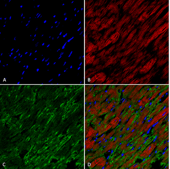
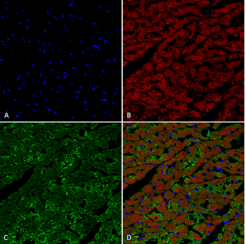
Immunohistochemistry analysis using Mouse Anti-Hexanoyl-Lysine adduct Monoclonal Antibody, Clone 5E8. Tissue: Heart. Species: Rat. Fixation: Formalin fixed, paraffin embedded. Primary Antibody: Mouse Anti-Hexanoyl-Lysine adduct Monoclonal Antibody at 1:26 for 2 hour at RT. Secondary Antibody: Goat Anti-Mouse IgG: Alexa Fluor 489. Counterstain: DAPI (blue) nuclear stain. Magnification: 63X. (A) DAPI (blue) nuclear stain. (B) Actin (C) Hexanoyl-Lysine adduct Antibody (D) Composite.
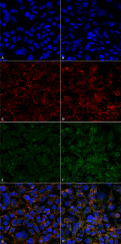
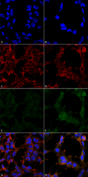
Immunocytochemistry/Immunofluorescence analysis using Mouse Anti-Hexanoyl-Lysine adduct Monoclonal Antibody, Clone 5E8. Tissue: Embryonic kidney epithelial cell line (HEK293). Species: Human. Fixation: 5% Formaldehyde for 5 min. Primary Antibody: Mouse Anti-Hexanoyl-Lysine adduct Monoclonal Antibody at 1:50 for 30-60 min at RT. Secondary Antibody: Goat Anti-Mouse Alexa Fluor 488 at 1:1500 for 30-60 min at RT. Counterstain: Phalloidin Alexa Fluor 633 F-Actin stain; DAPI (blue) nuclear stain at 1:250, 1:50000 for 30-60 min at RT. Magnification: 20X (2X Zoom). (A, C, E, G) - Untreated. (B, D, F, H) - Cells cultured overnight with 50 μM H2O2. (A, B) DAPI (blue) nuclear stain. (C, D) Phalloidin Alexa Fluor 633 F-Actin stain. (E, F) Hexanoyl-Lysine adduct Antibody. (G, H) Composite.
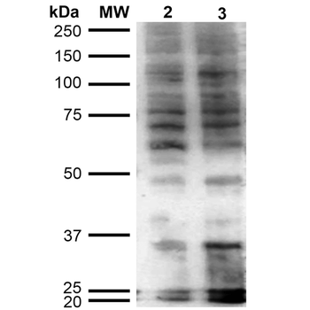
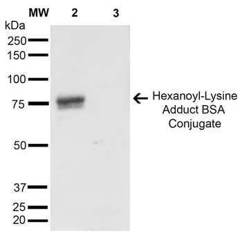
Western Blot analysis of Hexanoyl Lysine-BSA Conjugate showing detection of 67 kDa Hexanoyl-Lysine adduct protein using Mouse Anti-Hexanoyl-Lysine adduct Monoclonal Antibody, Clone 5E8. Lane 1: Molecular Weight Ladder (MW). Lane 2: Hexanoyl Lysine-BSA. Lane 3: BSA. Load: 0.5 μg. Block: 5% Skim Milk in TBST. Primary Antibody: Mouse Anti-Hexanoyl-Lysine adduct Monoclonal Antibody at 1:1000 for 2 hours at RT. Secondary Antibody: Goat Anti-Mouse IgG: HRP at 1:2000 for 60 min at RT. Color Development: ECL solution for 5 min in RT. Predicted/Observed Size: 67 kDa.
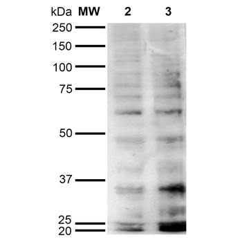
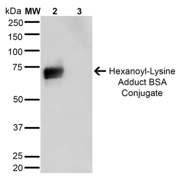
Western Blot analysis of Human Cervical cancer cell line (HeLa) lysate showing detection of Hexanoyl-Lysine adduct protein using Mouse Anti-Hexanoyl-Lysine adduct Monoclonal Antibody, Clone 5E8. Lane 1: Molecular Weight Ladder (MW). Lane 2: HeLa cell lysate. Lane 3: H2O2 treated HeLa cell lysate. Load: 12 μg. Block: 5% Skim Milk in TBST. Primary Antibody: Mouse Anti-Hexanoyl-Lysine adduct Monoclonal Antibody at 1:1000 for 2 hours at RT. Secondary Antibody: Goat Anti-Mouse IgG: HRP at 1:2000 for 60 min at RT. Color Development: ECL solution for 5 min in RT.
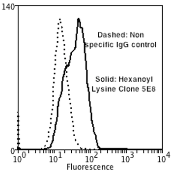
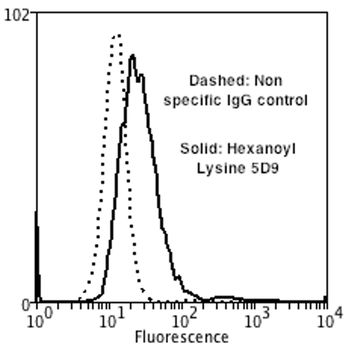
Flow Cytometry analysis using Mouse Anti-Hexanoyl-Lysine adduct Monoclonal Antibody, Clone 5E8. Tissue: Neuroblastoma cells (SH-SY5Y). Species: Human. Fixation: 90% Methanol. Primary Antibody: Mouse Anti-Hexanoyl-Lysine adduct Monoclonal Antibody at 1:50 for 30 min on ice. Secondary Antibody: Goat Anti-Mouse: PE at 1:100 for 20 min at RT. Isotype Control: Non Specific IgG.
Quick Database Links
Gene Symbol
Hexanoyl-Lysine adduct
Documents Download
Datasheet
Product Information
Request a Document
Protocol Information
WB
Western Blot (IB, immunoblot)
FACS
Fluorescence-Activated Cell Sorting (FC, Flow cytometry)
FC
Flow Cytometry
IF
Immunofluorescence
ICC
Immunocytochemistry
ELISA
Enzyme-linked Immunosorbent Assay (EIA)
Hexanoyl-Lysine adduct Antibody (Biotin) (orb396561)

- 0.0
Based on 0 reviews
Participating in our Biorbyt product reviews program enables you to support fellow scientists by sharing your firsthand experience with our products.
Login to Submit a ReviewAvailable Sizes
Select a size below
Choose Conjugation or Carrier Free Version
Free Secondary Antibody (20 ul)0/0
Please add an antibody product to your cart first.