You have no items in your shopping cart.
Description
Research Area
Pharmacology & Drug Discovery
Images & Validation
−Item 1 of 6
| Tested Applications | ELISA, FACS, FC, ICC, IF, IHC, WB |
|---|---|
| Dilution Range | WB (1:1000); ICC/IF (1:50); ELISA (1:1000); FACS (1:50); FCM (1:50); IHC (1:100) |
| Reactivity | All |
| Application Notes |
Key Properties
−| Host | Mouse |
|---|---|
| Clonality | Monoclonal |
| Isotype | IgG1 |
| Clone No. | 5D9 |
| Immunogen | Synthetic Hexanoyl modified Keyhole Limpet Hemocyanin (KLH). |
| Target | Hexanoyl-Lysine adduct |
| Purification | Protein G Purified |
| Conjugation | Biotin |
Storage & Handling
−| Storage | Conjugated antibodies should be stored according to the product label |
|---|---|
| Buffer/Preservatives | 136.36mM Ethanolamine, 133.23 mM Chlorides, 9.55mM Phosphates, 9.55mM Sodium Bicarbonate |
| Concentration | 1 mg/ml |
| Expiration Date | 12 months from date of receipt. |
| Disclaimer | For research use only |
Alternative Names
−Hexanoyl-Lysine adduct, HEL (Hexanoyl-Lysine adduct), HEL, HEL Adduct, Hexanoyl-Lys adduct, Hexanoyl-Lys, Hexanoyl-Lysine (HEL) adduct, Hexanoyl-Lys (HEL), Hexanoyl Lysine adduct, Hexanoyl-Lysine adduct-modified protein, Hexanoyl-Lysine Adduct (HEL)
Similar Products
−- Item 1 of 5
Hexanoyl-Lysine adduct Antibody (Biotin) [orb396561]
ELISA, FACS, FC, ICC, IF, WB
All
Mouse
Monoclonal
Biotin
100 μg
Quality Guarantee
Explore bioreagents carefree to elevate your research. All our products are rigorously tested for performance. If a product does not perform as described on its datasheet, our scientific support team will provide expert troubleshooting, a prompt replacement, or a refund. For full details, please see our Terms & Conditions and Buying Guide. Contact us at [email protected].

Immunohistochemistry analysis using Mouse Anti-Hexanoyl-Lysine adduct Monoclonal Antibody, Clone 5D9. Tissue: Kidney. Species: Rat. Primary Antibody: Mouse Anti-Hexanoyl-Lysine adduct Monoclonal Antibody at 1:100 for Overnight at 4°C, then 30 min at 37°C. Secondary Antibody: Goat Anti-Mouse IgG (H+L): FITC for 45 min at 37°C. Counterstain: DAPI for 3 min at RT. Magnification: 10X.
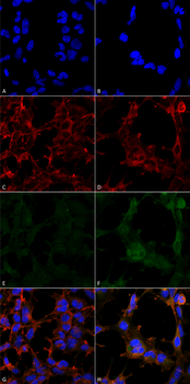
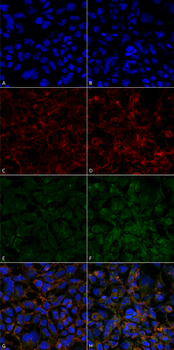
Immunocytochemistry/Immunofluorescence analysis using Mouse Anti-Hexanoyl-Lysine adduct Monoclonal Antibody, Clone 5D9. Tissue: Embryonic kidney epithelial cell line (HEK293). Species: Human. Fixation: 5% Formaldehyde for 5 min. Primary Antibody: Mouse Anti-Hexanoyl-Lysine adduct Monoclonal Antibody at 1:50 for 30-60 min at RT. Secondary Antibody: Goat Anti-Mouse Alexa Fluor 488 at 1:1500 for 30-60 min at RT. Counterstain: Phalloidin Alexa Fluor 633 F-Actin stain; DAPI (blue) nuclear stain at 1:250, 1:50000 for 30-60 min at RT. Magnification: 20X (2X Zoom). (A, C, E, G) - Untreated. (B, D, F, H) - Cells cultured overnight with 50 μM H2O2. (A, B) DAPI (blue) nuclear stain. (C, D) Phalloidin Alexa Fluor 633 F-Actin stain. (E, F) Hexanoyl-Lysine adduct Antibody. (G, H) Composite.
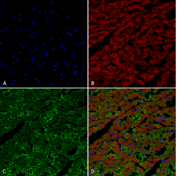
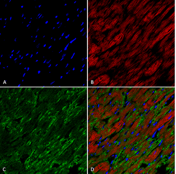
Immunohistochemistry analysis using Mouse Anti-Hexanoyl-Lysine adduct Monoclonal Antibody, Clone 5D9. Tissue: Heart. Species: Rat. Fixation: Formalin fixed, paraffin embedded. Primary Antibody: Mouse Anti-Hexanoyl-Lysine adduct Monoclonal Antibody at 1:25 for 1 hour at RT. Secondary Antibody: Goat Anti-Mouse IgG: Alexa Fluor 488. Counterstain: DAPI (blue) nuclear stain. Magnification: 63X. (A) DAPI (blue) nuclear stain. (B) Actin (C) Hexanoyl-Lysine adduct Antibody (D) Composite.
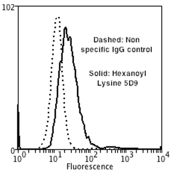
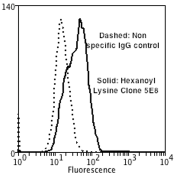
Flow Cytometry analysis using Mouse Anti-Hexanoyl-Lysine adduct Monoclonal Antibody, Clone 5D9. Tissue: Neuroblastoma cells (SH-SY5Y). Species: Human. Fixation: 90% Methanol. Primary Antibody: Mouse Anti-Hexanoyl-Lysine adduct Monoclonal Antibody at 1:50 for 30 min on ice. Secondary Antibody: Goat Anti-Mouse: PE at 1:100 for 20 min at RT. Isotype Control: Non Specific IgG. Cells were subject to oxidative stress by treating with 250 μM H2O2 for 24 hours.
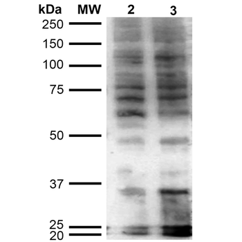
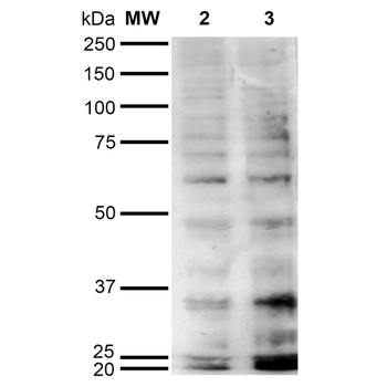
Western Blot analysis of Human Cervical cancer cell line (HeLa) lysate showing detection of Hexanoyl-Lysine adduct protein using Mouse Anti-Hexanoyl-Lysine adduct Monoclonal Antibody, Clone 5D9. Lane 1: Molecular Weight Ladder (MW). Lane 2: HeLa cell lysate. Lane 3: H2O2 treated HeLa cell lysate. Load: 12 μg. Block: 5% Skim Milk in TBST. Primary Antibody: Mouse Anti-Hexanoyl-Lysine adduct Monoclonal Antibody at 1:1000 for 2 hours at RT. Secondary Antibody: Goat Anti-Mouse IgG: HRP at 1:2000 for 60 min at RT. Color Development: ECL solution for 5 min in RT.
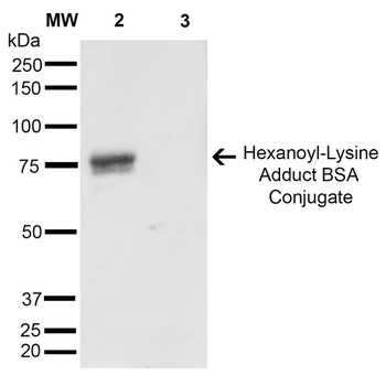
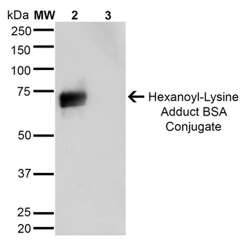
Western Blot analysis of Hexanoyl Lysine-BSA Conjugate showing detection of 67 kDa Hexanoyl-Lysine adduct protein using Mouse Anti-Hexanoyl-Lysine adduct Monoclonal Antibody, Clone 5D9. Lane 1: Molecular Weight Ladder (MW). Lane 2: Hexanoyl Lysine-BSA. Lane 3: BSA. Load: 0.5 μg. Block: 5% Skim Milk in TBST. Primary Antibody: Mouse Anti-Hexanoyl-Lysine adduct Monoclonal Antibody at 1:1000 for 2 hours at RT. Secondary Antibody: Goat Anti-Mouse IgG: HRP at 1:2000 for 60 min at RT. Color Development: ECL solution for 5 min in RT. Predicted/Observed Size: 67 kDa.
Quick Database Links
Gene Symbol
Hexanoyl-Lysine adduct
Documents Download
Datasheet
Product Information
Request a Document
Protocol Information
WB
Western Blot (IB, immunoblot)
IHC
Immunohistochemistry
FACS
Fluorescence-Activated Cell Sorting (FC, Flow cytometry)
FC
Flow Cytometry
IF
Immunofluorescence
ICC
Immunocytochemistry
ELISA
Enzyme-linked Immunosorbent Assay (EIA)
Hexanoyl-Lysine adduct Antibody (Biotin) (orb396543)

- 0.0
Based on 0 reviews
Participating in our Biorbyt product reviews program enables you to support fellow scientists by sharing your firsthand experience with our products.
Login to Submit a ReviewAvailable Sizes
Select a size below
Choose Conjugation or Carrier Free Version
Free Secondary Antibody (20 ul)0/0
Please add an antibody product to your cart first.