You have no items in your shopping cart.
Description
Images & Validation
−Item 1 of 3
| Tested Applications | ELISA, IF, WB |
|---|---|
| Dilution Range | ELISA: 1:128000, WB: 1-3ug/ml, IHC-P: 10ug/ml |
| Reactivity | Human, Mouse |
| Predicted Reactivity | Canine, Rat |
| Application Notes |
Key Properties
−| Host | Goat |
|---|---|
| Clonality | Polyclonal |
| Target | IFT88 / Polaris |
| Protein Sequence | KKRIDEDDFADEE |
| Molecular Weight | 94.3; 93.2 |
| Purification | Purified from goat serum by ammonium sulphate precipitation followed by antigen affinity chromatography using the immunizing peptide. |
| Conjugation | Unconjugated |
Storage & Handling
−| Storage | Maintain refrigerated at 2-8°C for up to 2 weeks. For long term storage store at -20°C in small aliquots to prevent freeze-thaw cycles. |
|---|---|
| Buffer/Preservatives | Supplied at 0.5 mg/ml in Tris saline, 0.02% sodium azide, pH 7.3 with 0.5% bovine serum albumin. Aliquot and store at -20°C. Minimize freezing and thawing. |
| Expiration Date | 12 months from date of receipt. |
| Disclaimer | For research use only |
Alternative Names
−anti IFT88 antibody, anti polaris antibody, anti TTC10 antibody, anti tetratricopeptide repeat domain 10 antibody, anti HGNC:20606 antibody, anti D13S1056E antibody, anti DAF19 antibody, anti IFT88 antibody, anti MGC26259 antibody, anti RP11-172H24.2 antibody, anti TG737 antibody, anti hTg737 antibody, anti TPR repeat protein 10 antibody, anti Probe hTg737 (polycystic kidney disease, autosomal recessive) antibody, anti Tg737 protein antibody, anti intraflagellar transport 88 homolog antibody, anti recessive polycystic kidney disease protein Tg737 homolog antibody, anti intraflagellar transport 88 homolog (Chlamydomonas) antibody
Similar Products
−- Item 1 of 5
IFT88 Antibody [orb1247371]
ELISA, FC, IF, IHC, WB
Canine, Rat
Human, Mouse
Goat
Polyclonal
Unconjugated
0.1 mg

Quality Guarantee
Explore bioreagents carefree to elevate your research. All our products are rigorously tested for performance. If a product does not perform as described on its datasheet, our scientific support team will provide expert troubleshooting, a prompt replacement, or a refund. For full details, please see our Terms & Conditions and Buying Guide. Contact us at [email protected].

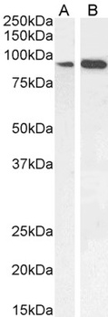
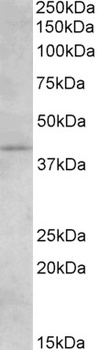
2 µg/ml staining of K562 (A), and 3 ug/ml THP-1 (B), U251 (C) and KNRK (D) cell lysate (35 µg protein in RIPA buffer). Detected by chemiluminescence.
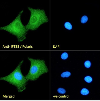
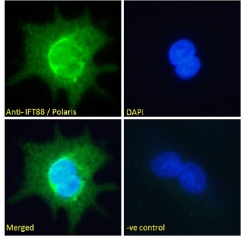
Immunofluorescence analysis of paraformaldehyde fixed A549 cells, permeabilized with 0.15% Triton. Primary incubation 1hr (10 ug/ml) followed by Alexa Fluor 488 secondary antibody (2 ug/ml), showing cytoplasmic staining. The nuclear stain is DAPI (blue). Negative control: Unimmunized goat IgG (10 ug/ml) followed by Alexa Fluor 488 secondary antibody (2 ug/ml).
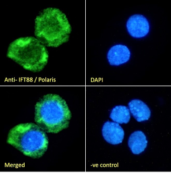
Immunofluorescence analysis of paraformaldehyde fixed HepG2 cells, permeabilized with 0.15% Triton. Primary incubation 1hr (10 ug/ml) followed by Alexa Fluor 488 secondary antibody (2 ug/ml), showing cytoplasmic staining. The nuclear stain is DAPI (blue). Negative control: Unimmunized goat IgG (10 ug/ml) followed by Alexa Fluor 488 secondary antibody (2 ug/ml).
Documents Download
Datasheet
Product Information
Request a Document
Protocol Information
WB
Western Blot (IB, immunoblot)
IF
Immunofluorescence
ELISA
Enzyme-linked Immunosorbent Assay (EIA)
IFT88/Polaris Antibody (orb19151)

- 0.0
Based on 0 reviews
Participating in our Biorbyt product reviews program enables you to support fellow scientists by sharing your firsthand experience with our products.
Login to Submit a ReviewAvailable Sizes
Select a size below
Choose Conjugation or Carrier Free Version
Free Secondary Antibody (20 ul)0/0
Please add an antibody product to your cart first.