You have no items in your shopping cart.
Description
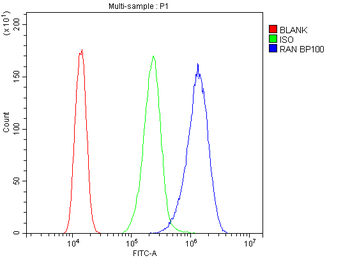
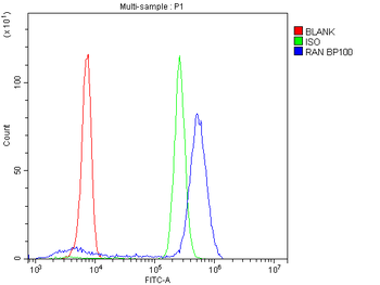
Research Area
Epigenetics, Neuroscience
Images & Validation
−Item 1 of 7
| Tested Applications | ChIP, ELISA, IF, IHC, WB |
|---|---|
| Dilution Range | ELISA: 1:64000, WB: 0.1-0.5 μg/ml, IHC-P: 2-3 μg/ml |
| Reactivity | Human |
| Predicted Reactivity | Bovine, Canine, Mouse, Porcine, Rat |
| Application Notes |
Key Properties
−| Clonality | Polyclonal |
|---|---|
| Immunogen | Peptide with sequence EVQLGLGRVYPRPPSC, from the N Terminus of the protein sequence |
| Target | Androgen Receptor |
| Protein Sequence | EVQLGLGRVYPRPPSC |
| Molecular Weight | Approx 100kDa band observed in Human Cerebellum lysates (calculated MW of 99.2kDa) |
| Purity | Purified from goat serum by ammonium sulphate precipitation followed by antigen affinity chromatography using the immunizing peptide |
| Purification | Purified from goat serum by ammonium sulphate precipitation followed by antigen affinity chromatography using the immunizing peptide. |
| Conjugation | Unconjugated |
Storage & Handling
−| Storage | Maintain refrigerated at 2-8°C for up to 2 weeks. For long term storage store at -20°C in small aliquots to prevent freeze-thaw cycles. |
|---|---|
| Form/Appearance | Liquid |
| Buffer/Preservatives | 0.5 mg/ml in Tris saline, 0.02% sodium azide, pH7.3 with 0.5% bovine serum albumin |
| Expiration Date | 12 months from date of receipt. |
| Disclaimer | For research use only |
Alternative Names
−anti androgen receptor antibody, anti dihydrotestosterone receptor antibody, anti AR antibody, anti KD antibody, anti AIS antibody, anti TFM antibody, anti DHTR antibody, anti SBMA antibody, anti NR3C4 antibody, anti SMAX1 antibody, anti HUMARA antibody, anti HYSP1 antibody, anti androgen receptor antibody, anti dihydrotestosterone receptor antibody, anti RP11-383C12.1 antibody
Similar Products
−- Item 1 of 10
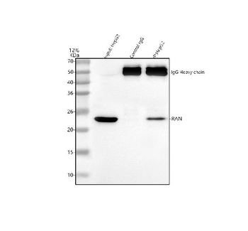
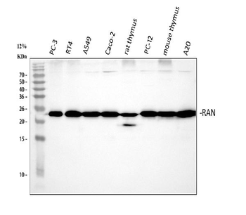
Ran Rabbit Polyclonal Antibody [orb402333]
FC, ICC, IF, IHC, IP, WB
Human, Mouse, Rat
Rabbit
Polyclonal
Unconjugated
100 μg - Item 1 of 7
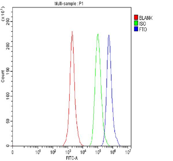
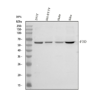
FTO Rabbit Polyclonal Antibody [orb763045]
ELISA, FC, ICC, IF, IHC, WB
Human, Mouse, Rat
Rabbit
Polyclonal
Unconjugated
100 μg - Item 1 of 4
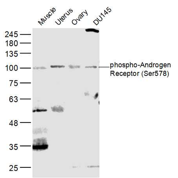
Phospho-AR/Androgen receptor (Ser578) Rabbit Polyclonal Antibody [orb10129]
FC, IF, IHC-Fr, IHC-P, WB
Bovine, Canine, Equine, Gallus, Porcine
Human, Mouse, Rat
Rabbit
Polyclonal
Unconjugated
50 μl, 100 μl, 200 μl - Item 1 of 6
Phospho-AR/Androgen receptor (Ser650) Rabbit Polyclonal Antibody [orb4542]
FC, IF, IHC-Fr, IHC-P
Canine, Porcine, Rabbit
Human, Mouse, Rat
Rabbit
Polyclonal
Unconjugated
50 μl, 100 μl, 200 μl - Item 1 of 6
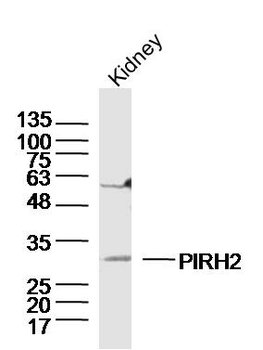
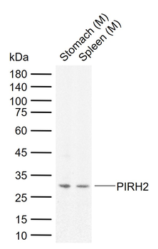
PIRH2 Rabbit Polyclonal Antibody [orb2868]
FC, ICC, IF, IHC-Fr, IHC-P, WB
Bovine, Canine, Equine, Porcine, Rat, Sheep
Human, Mouse
Rabbit
Polyclonal
Unconjugated
50 μl, 100 μl, 200 μl

Quality Guarantee
Explore bioreagents carefree to elevate your research. All our products are rigorously tested for performance. If a product does not perform as described on its datasheet, our scientific support team will provide expert troubleshooting, a prompt replacement, or a refund. For full details, please see our Terms & Conditions and Buying Guide. Contact us at [email protected].
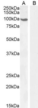
staining of Human Cerebellum (A) and (0.1 μg/ml) negative control Pancreas (B) lysate (35 μg protein in RIPA buffer). Detected by chemiluminescence.
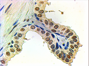
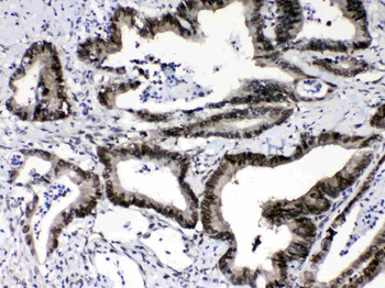
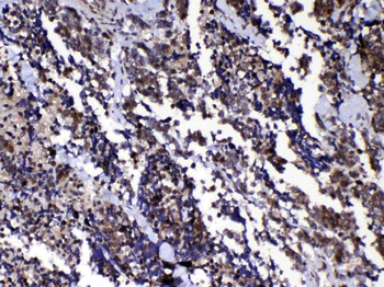
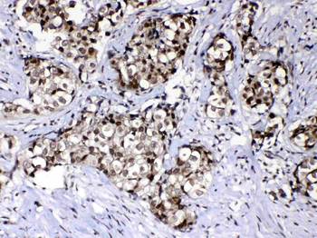
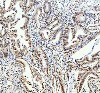
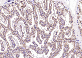
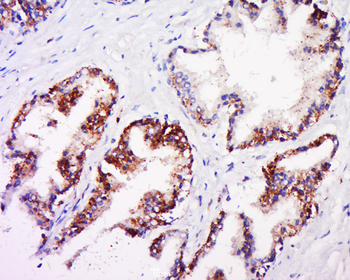
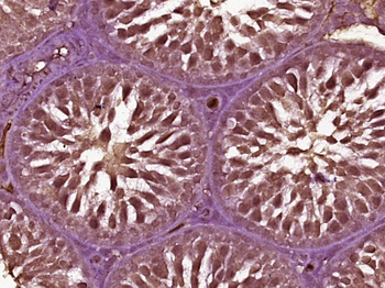
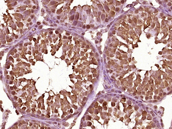
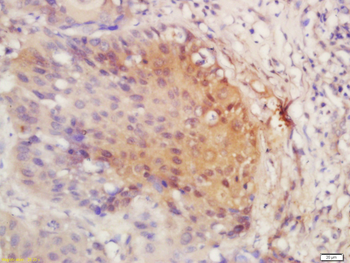
2 μg/ml staining of paraffin embedded Human Prostate. Steamed antigen retrieval with citrate buffer pH 6, AP-staining.
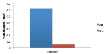
ChIP of 2 μg with 1 μg DHT-treated HEC50 chromatin using the spin column sonication kit (Protein G) measuring FKBP5 enrichment.
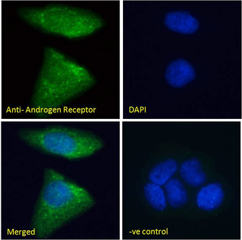
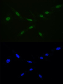
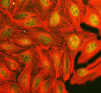
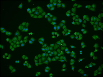
Immunofluorescence analysis of paraformaldehyde fixed MCF7 cells, permeabilized with 0.15% Triton. Primary incubation 1hr (10 μg/ml) followed by Alexa Fluor 488 secondary antibody (2 μg/ml), showing Mitochondrial/cytoplasmic staining. The nuclear stain is DAPI (blue). Negative control: Unimmunized goat IgG (10 μg/ml) followed by Alexa Fluor 488 secondary antibody (2 μg/ml).
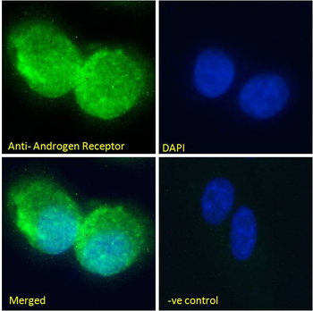
Immunofluorescence analysis of paraformaldehyde fixed U2OS cells, permeabilized with 0.15% Triton. Primary incubation 1hr (10 μg/ml) followed by Alexa Fluor 488 secondary antibody (2 μg/ml), showing Mitochondrial/cytoplasmic staining. The nuclear stain is DAPI (blue). Negative control: Unimmunized goat IgG (10 μg/ml) followed by Alexa Fluor 488 secondary antibody (2 μg/ml).
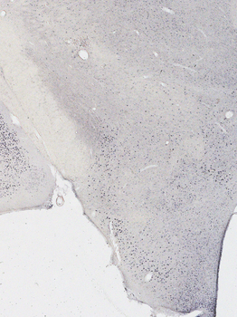
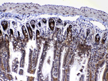
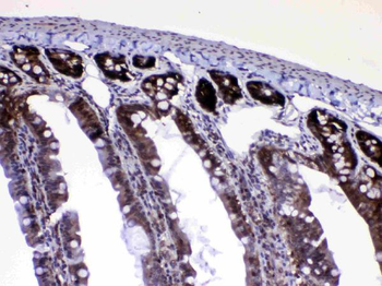
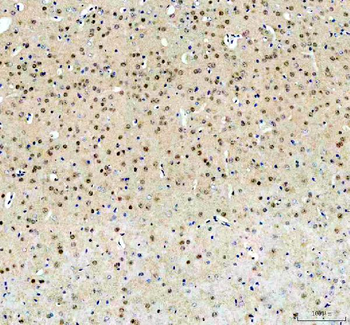
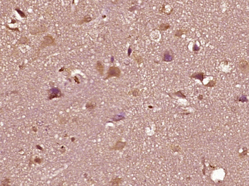
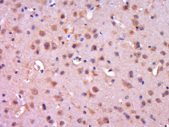
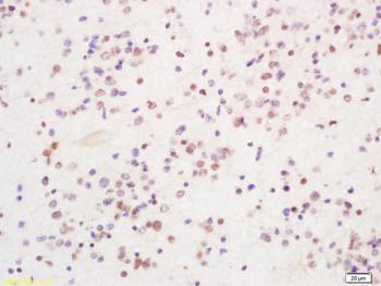
Immunostaining of 25μm thick cryosections of PFA-perfused Mouse Hypothalamus, antigen retrieval with citrate buffer Ph 6 at 80°C for 30 min, HRP-staining with Ni-DAB after Biotin-SP-antigoat amplification.

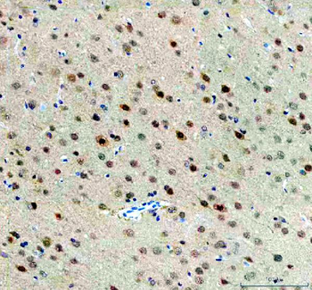
Immunostaining of 25μm thick cryosections of PFA-perfused Mouse Amygdala, antigen retrieval with citrate buffer Ph 6 at 80°C for 30 min, HRP-staining with Ni-DAB after Biotin-SP-antigoat amplification.
Documents Download
Datasheet
Product Information
Request a Document
Protocol Information
WB
Western Blot (IB, immunoblot)
IHC
Immunohistochemistry
IF
Immunofluorescence
ELISA
Enzyme-linked Immunosorbent Assay (EIA)
ChIP
Chromatin Immunoprecipitation
Filter by Applications
Filter by Species
Xueying Wang et al. Chronic exposure to F-53B disrupts sperm quality and steroidogenic regulation in male rats Toxicol Lett, 419, 111885 (2026)
Applications
WB
Reactivity
Rat
Androgen Receptor Antibody (orb18847)

- 0.0
Based on 0 reviews
Participating in our Biorbyt product reviews program enables you to support fellow scientists by sharing your firsthand experience with our products.
Login to Submit a ReviewAvailable Sizes
Select a size below
Choose Conjugation or Carrier Free Version
Free Secondary Antibody (20 ul)0/0
Please add an antibody product to your cart first.