You have no items in your shopping cart.
GLUD1 Antibody
SKU: orb750578
Description
Images & Validation
−Item 1 of 3
| Tested Applications | ELISA, IP, WB |
|---|---|
| Dilution Range | ELISA: 1:4,000 - 1:16,000, IP: 1:100, WB: 1:1,000 - 1:3,000 |
| Reactivity | Bovine |
| Application Notes |
Key Properties
−| Antibody Type | Primary Antibody |
|---|---|
| Host | Rabbit |
| Clonality | Polyclonal |
| Isotype | Antiserum |
| Immunogen | This antibody was prepared from whole rabbit serum produced by repeated immunizations with a full length Glutamate Dehydrogenase protein isolated from Bovine Liver. |
| Target | GLUD1 |
| Purity | This product was prepared from monospecific antiserum by a delipidation and defibrination. Assay by immunoelectrophoresis resulted in a single precipitin arc against anti-rabbit serum, purified and partially purified Glutamate Dehydrogenase [Bovine Liver]. BLAST analysis was used to determine that cross reactivity is suggested for both mitochondrial and brain isoforms (GDH1 and GDH2), from both bovine and human sources. Additionally similar reactivity is suggested for most primate species including green monkey, white gibbon, chimpanzee orangutan, and gorilla. A high degree of sequence homology is also noted for GDH from chicken, mouse, rat, dog, and other mammals as well as Xenopus tropicalis, zebrafish, rainbow trout and Atlantic salmon. Cross reactivity against Glutamate Dehydrogenase from other tissues and species may occur but have not been specifically determined. |
| Conjugation | Unconjugated |
Storage & Handling
−| Storage | Store vial at 4° C prior to restoration. For extended storage aliquot contents and freeze at -20° C or below. Avoid cycles of freezing and thawing. Centrifuge product if not completely clear after standing at room temperature. This product is stable for several weeks at 4° C as an undiluted liquid. Dilute only prior to immediate use. |
|---|---|
| Form/Appearance | Lyophilized |
| Buffer/Preservatives | 0.01% (w/v) Sodium Azide |
| Concentration | 85 mg/mL |
| Expiration Date | 12 months from date of receipt. |
| Disclaimer | For research use only |
Alternative Names
−rabbit anti-Glutamate Dehydrogenase Antibody, Glutamate dehydrogenase 1 mitochondrial, GDH 1
Similar Products
−- Item 1 of 16
GLUD1 Rabbit Polyclonal Antibody [orb579551]
IHC, WB
Bovine, Canine, Equine, Guinea pig, Mouse, Porcine, Rabbit, Sheep, Zebrafish
Human, Rat
Rabbit
Polyclonal
Unconjugated
100 μl - Item 1 of 15
GLUD1/2 Rabbit Polyclonal Antibody [orb1152344]
ELISA, FC, ICC, IF, IHC, WB
Human, Mouse, Rat
Rabbit
Polyclonal
Unconjugated
100 μg - Item 1 of 8
- Item 1 of 5
Glutamate Dehydrogenase Antibody [orb20463]
ELISA, IF, IHC, WB
Bovine, Canine
Human, Mouse, Rat
Goat
Polyclonal
Unconjugated
100 μg - Item 1 of 1
Human Glutamate Dehydrogenase 1 (GDH) ELISA Kit [orb775791]
Human
0.16-10 ng/mL
0.064 ng/mL
96 T, 48 T


Quality Guarantee
Explore bioreagents carefree to elevate your research. All our products are rigorously tested for performance. If a product does not perform as described on its datasheet, our scientific support team will provide expert troubleshooting, a prompt replacement, or a refund. For full details, please see our Terms & Conditions and Buying Guide. Contact us at [email protected].
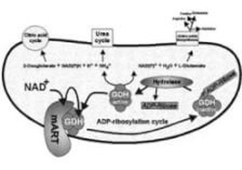
Metabolic pathways that may be affected by the inhibition of GDH are indicated. mART, mitochondrial ADP-ribosyl transferase.
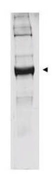
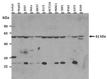
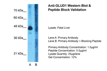
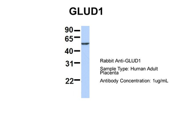
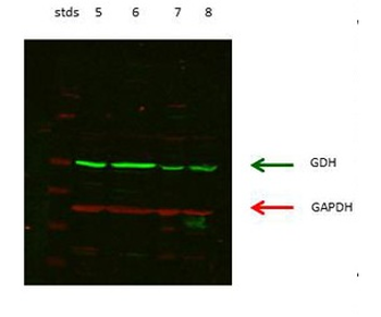
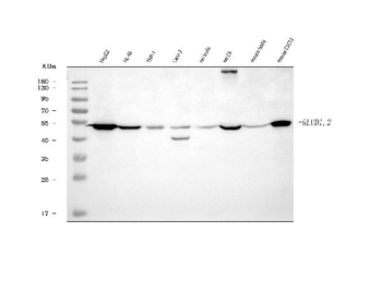
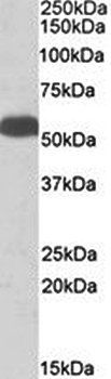
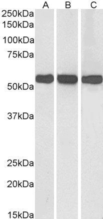
Western blot analysis is shown using Biorbyt's anti-bovine glutamate dehydrogenase antibody to detect the enzyme from bovine liver preparations. Comparison to a molecular weight marker indicates a predominant band of ~62 kDa. The higher molecular weight band may represent a subunit dimer. A 4-20% gradient gel was used to separate proteins prior to transfer to 0.2 µm nitrocellulose. The blot was incubated with a 1:1000 dilution of the antibody for 2 h at room temperature followed by detection using IRDye™800 labeled Goat-a-Rabbit IgG [H&L] diluted 1:5000 for 45 min at room temperature.
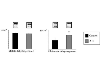
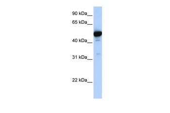
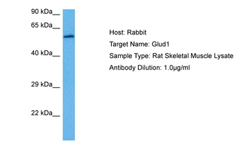
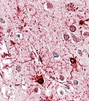
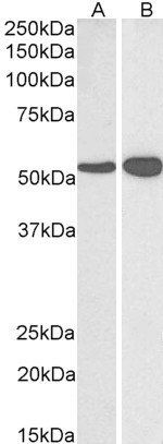
Western Blot using Anti-Glutamate Dehydrogenase (Bovine Liver) (RABBIT) Antibody. Two different molecular weight groups of isoforms were detected for MDH1 at about 36 kDa using Anti-MDH1 (pig heart) orb750614 at 1:1500, Fluorescent Cy-5 labelled secondary at 1:500 and for GDH at about 60 kDa using Anti-GDH (bovine liver) orb750578 at 1:1500, anti-rabbit secondary at 1:500. Light intensities revealed unchanged amount of total soluble MDH1 but increased amount of total soluble GDH in AD when compared to controls. Values of light intensities are given as mean ± S.E.M. *p ≤ 0.05.
Documents Download
Datasheet
Product Information
Request a Document
Protocol Information
WB
Western Blot (IB, immunoblot)
ELISA
Enzyme-linked Immunosorbent Assay (EIA)
IP
Immunoprecipitation
GLUD1 Antibody (orb750578)

- 0.0
Based on 0 reviews
Participating in our Biorbyt product reviews program enables you to support fellow scientists by sharing your firsthand experience with our products.
Login to Submit a ReviewAvailable Sizes
Select a size below