You have no items in your shopping cart.
Description
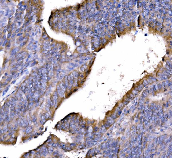
Research Area
Signal Transduction
Images & Validation
−Item 1 of 3
| Tested Applications | ELISA, IP, WB |
|---|---|
| Dilution Range | ELISA: 1:10,000 - 1:50,000, IP: 1:100, WB: 1:500 - 1:3,000 |
| Reactivity | Human, Primate |
| Application Notes |
Key Properties
−| Antibody Type | Primary Antibody |
|---|---|
| Host | Rabbit |
| Clonality | Polyclonal |
| Isotype | IgG |
| Immunogen | This affinity-purified antibody was prepared from whole rabbit serum produced by repeated immunizations with a synthetic peptide corresponding to an internal region near amino acids 400-425 of Human GGA3. |
| Target | GGA3 |
| Purity | This affinity purified antibody is directed against human GGA3 protein. The product was affinity purified from monospecific antiserum by immunoaffinity purification. A BLAST analysis was used to suggest reactivity with this protein from human and chimpanzee based on 100% homology for the immunogen sequence. Cross reactivity may occur with GGA3 from macaque (93% homology) and orangutan (87% homology) sources. The immunizing sequence is present on both the long (isoform 1) and short (isoform 2) transcript variants for this protein. |
| Conjugation | Unconjugated |
Storage & Handling
−| Storage | Store vial at -20° C prior to opening. Aliquot contents and freeze at -20° C or below for extended storage. Avoid cycles of freezing and thawing. Centrifuge product if not completely clear after standing at room temperature. This product is stable for several weeks at 4° C as an undiluted liquid. Dilute only prior to immediate use. |
|---|---|
| Form/Appearance | Liquid (sterile filtered) |
| Buffer/Preservatives | Preservative: 0.01% (w/v) Sodium Azide. Stabilizer: None; Buffer: 0.02 M Potassium Phosphate, 0.15 M Sodium Chloride, pH 7.2 |
| Concentration | 1.0mg/mL |
| Expiration Date | 12 months from date of receipt. |
| Dry Ice Shipping | Please note: This product requires shipment on dry ice. A dry ice surcharge will apply. |
| Disclaimer | For research use only |
Alternative Names
−rabbit anti-GGA3 antibody, GGA-3, GGA 3, ADP-ribosylation factor-binding protein GGA3, Golgi-localized gamma ear-containing ARF-binding protein 3
Similar Products
−- Item 1 of 6
GGA3 Rabbit Polyclonal Antibody [orb763170]
ELISA, FC, ICC, IF, IHC, WB
Human
Rabbit
Polyclonal
Unconjugated
100 μg - Item 1 of 1
- Item 1 of 1
- Item 1 of 1
Gamma-Tubulin GGA3 Mouse Monoclonal Antibody [orb18289]
ICC, WB
Human, Mouse, Rat
Mouse
Monoclonal
Unconjugated
100 μg - Item 1 of 1
GGA3 Rabbit Polyclonal Antibody [orb330809]
WB
Bovine, Canine, Equine, Guinea pig, Mouse, Rabbit, Rat, Zebrafish
Human
Rabbit
Polyclonal
Unconjugated
100 μl

Quality Guarantee
Explore bioreagents carefree to elevate your research. All our products are rigorously tested for performance. If a product does not perform as described on its datasheet, our scientific support team will provide expert troubleshooting, a prompt replacement, or a refund. For full details, please see our Terms & Conditions and Buying Guide. Contact us at [email protected].
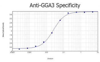
ELISA results of purified Rabbit anti-GGA3 Antibody tested against BSA-conjugated peptide of immunizing peptide. Each well was coated in duplicate with 0.1 µg of conjugate. The starting dilution of antibody was 5 µg/ml and the X-axis represents the Log10 of a 3-fold dilution. This titration is a 4-parameter curve fit where the IC50 is defined as the titer of the antibody. Assay performed using 3% fish gel, Goat anti-Rabbit IgG Antibody Peroxidase Conjugated (Min X Bv Ch Gt GP Ham Hs Hu Ms Rt & Sh Serum Proteins) (p/n orb347654) and TMB ELISA Peroxidase Substrate (p/n orb348651).
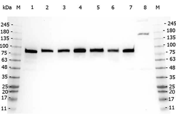
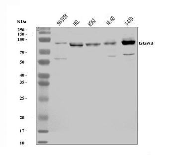
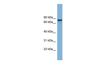
Western Blot of Rabbit anti-GGA3 antibody. Marker: Opal Pre-stained ladder. Lane 1: HEK293 lysate (p/n orb348669). Lane 2: HeLa Lysate (p/n orb348668). Lane 3: MCF-7 Lysate (p/n orb348664). Lane 4: Jurkat Lysate. Lane 5: A431 Lysate (p/n orb348665). Lane 6: LNCaP Lysate (p/n orb348694). Lane 7: A-172 Lysate (p/n orb348708). Lane 8: NIH/3T3 Lysate (p/n orb348714). Load: 35 µg per lane. Primary antibody: GGA3 antibody at 1:5000 for overnight at 4°C. Secondary antibody: Peroxidase rabbit secondary antibody (p/n orb347654) at 1:30000 for 60 min at RT. Blocking Buffer: 1% Casein-TTBS for 30 min at RT. Predicted/Observed size: 78 kDa for GGA3.
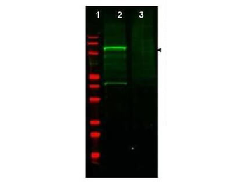
Western blot using Biorbyt's Affinity Purified anti-GGA3 antibody shows detection of a band at ~110 kDa corresponding to GFP-GGA3 fusion protein present in a lysate of HEK293 cells over- expressing the recombinant protein (lane 2, arrowhead). Pre-incubation of antibody with immunizing peptide blocks specific staining (lane 3). MW markers are shown in lane 1 (700 nm channel - red). Approximately 35 µg of lysate was separated on a 16% Tricine gel by SDS-PAGE and transferred onto nitrocellulose. After blocking the membrane was probed with the primary antibody diluted to 1:600. Reaction occurred overnight at 4°C followed by washes and reaction with a 1:10000 dilution of IRDye800 conjugated Gt-a-Rabbit IgG [H&L] for 45 min at room temperature (800 nm channel - green).
Documents Download
Datasheet
Product Information
Request a Document
Protocol Information
WB
Western Blot (IB, immunoblot)
ELISA
Enzyme-linked Immunosorbent Assay (EIA)
IP
Immunoprecipitation
GGA3 Antibody (orb345520)

- 0.0
Based on 0 reviews
Participating in our Biorbyt product reviews program enables you to support fellow scientists by sharing your firsthand experience with our products.
Login to Submit a ReviewAvailable Sizes
Select a size below
Choose Conjugation or Carrier Free Version
Free Secondary Antibody (20 ul)0/0
Please add an antibody product to your cart first.