You have no items in your shopping cart.
Description
Research Area
Cardiovascular Research
Images & Validation
−Item 1 of 5
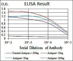
| Tested Applications | ELISA, IHC, IP, WB |
|---|---|
| Dilution Range | ELISA: 1:5,000 - 1:10,000, IHC: 1:50 - 1:200, IP: 1:100, WB: 1:5,000 - 1:10,000 |
| Reactivity | Human |
| Application Notes |
Key Properties
−| Antibody Type | Primary Antibody |
|---|---|
| Host | Rabbit |
| Clonality | Polyclonal |
| Isotype | IgG |
| Immunogen | Fibronectin was purified from Human plasma by binding to a denatured gelatin column followed by elution with high concentrations of arginine. The eluted material was further purified by gel filtration. Immunization occurred after single-band purity was assessed by SDS-PAGE. |
| Target | FN1 |
| Purity | This product has been prepared by immunoaffinity chromatography using immobilized antigens followed by extensive cross-adsorption against human serum proteins and collagen and non-collagen extracellular matrix proteins to remove any unwanted specificities. Typically less than 1% cross reactivity against other extracellular matrix proteins was detected by ELISA against purified standards. This antibody reacts with human Fibronectin and has negligible cross-reactivity with Type I, II, III, IV, V or VI Collagens or Laminin. Non-specific cross reaction of anti-Fibronectin antibodies with other human serum proteins or non-Fibronectin extracellular matrix proteins is negligible. |
| Conjugation | Unconjugated |
Storage & Handling
−| Storage | Store vial at 4° C prior to opening. This product is stable at 4° C as an undiluted liquid. Dilute only prior to immediate use. For extended storage mix with an equal volume of glycerol, aliquot contents and freeze at -20° C or below. Avoid cycles of freezing and thawing. |
|---|---|
| Form/Appearance | Liquid (sterile filtered) |
| Buffer/Preservatives | Preservative: 0.01% (w/v) Sodium Azide. Stabilizer: None; Buffer: 0.02 M Potassium Phosphate, 0.15 M Sodium Chloride, pH 7.2 |
| Concentration | 1.0 mg/mL |
| Expiration Date | 12 months from date of receipt. |
| Disclaimer | For research use only |
Alternative Names
−rabbit anti-Fibronectin antibody, FN1, FN, Cold-insoluble globulin, CIG, Anastellin, Ugl-Y1, Ugl-Y2, Ugl-Y3
Similar Products
−- Item 1 of 4
Fibronectin/FN1 Rabbit Polyclonal Antibody [orb10665]
IF, IHC-Fr, IHC-P
Bovine, Canine
Human, Mouse, Rat
Rabbit
Polyclonal
Unconjugated
50 μl, 100 μl, 200 μl - Item 1 of 6
- Item 1 of 4
FN1 rabbit pAb Antibody [orb765230]
ELISA, IF, IHC, WB
Human, Mouse, Rat
Polyclonal
Unconjugated
100 μl - Item 1 of 5
Fibronectin/FN1 Rabbit Polyclonal Antibody [orb570334]
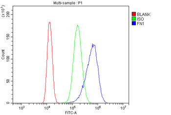
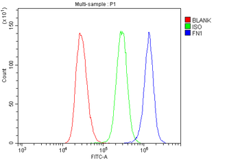
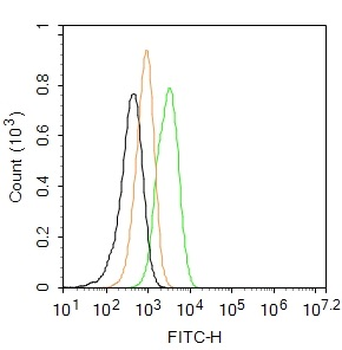
ELISA, FC, IHC, WB
Human
Rabbit
Polyclonal
Unconjugated
100 μg - Item 1 of 4
Fibronectin/Ugl-Y3 Rabbit Polyclonal Antibody (FITC) [orb222081]
ICC
Bovine, Canine, Equine, Gallus, Porcine, Rabbit
Human, Mouse, Rat
Rabbit
Polyclonal
FITC
100 μl


Quality Guarantee
Explore bioreagents carefree to elevate your research. All our products are rigorously tested for performance. If a product does not perform as described on its datasheet, our scientific support team will provide expert troubleshooting, a prompt replacement, or a refund. For full details, please see our Terms & Conditions and Buying Guide. Contact us at [email protected].
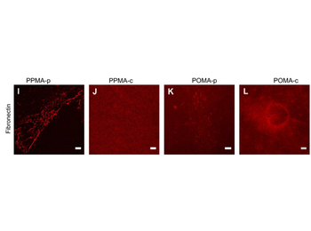
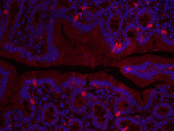
Immunofluorescence of Anti-Fibronectin Antibody. Representative images of isolated EC cultured on various MA-coated surfaces after 24 h exposure to 0.5 dyn/cm2 immunofluorescence-labelled for Fibronectin. A strong rearrangement of the initial Fibronectin layer into coarse fibrils (under venous shear stress) (Fig. 3I) with severe displacements of Fibronectin occurred on PPMA-p. Only slight Fibronectin reorganization into fine fibrils (PPMA-c Fig. 3J) or no Fibronectin reorganization at all (POMA-p and POMA-c Fig. 3K and L) were observed as expected from the higher Fibronectin anchorage strength to these latter substrates in comparison to PPMA-p. These findings are in line with earlier results at static cell culture conditions of isolated EC [4e6] showing the dependence of adhesion and stress fibre patterns on the matrix anchorage to the polymer surface, which were now attenuated by the application of shear stress. Scale bar: 10 mm.
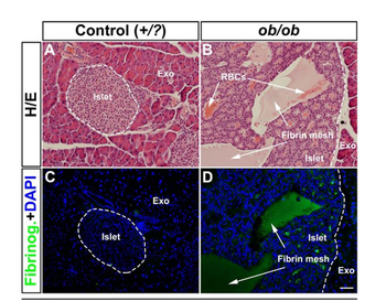
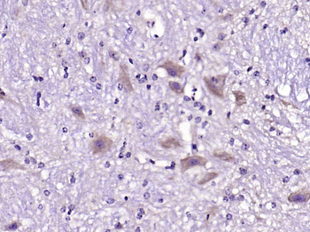
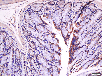
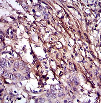
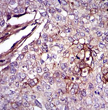
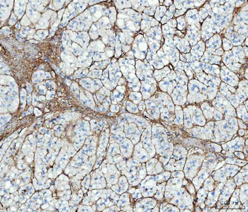
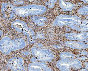
Immunohistochemistry of Anti-Fibronectin Antibody. Immunohistochemical assessment of proteins involved in blood coagulation in ob/ob pancreas. (A, B) Hematoxylin/Eosin staining of an islet from a lean control (A) and a ob/ob (B) pancreas at 52 weeks. Note the accumulation of RBCs (white arrows in (B). (C, D) Consecutive sections to (A, B) stained for Fibrinogen (green) and DAPI (Blue) indicating the presence of a fibrin mesh within the areas of the lesions (white arrows in (D) compare with (B)). Scale bar in (D) is 50 µm.
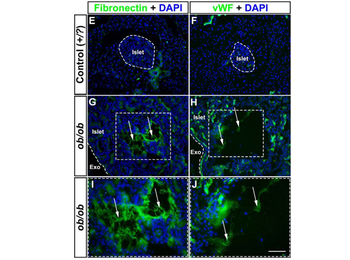
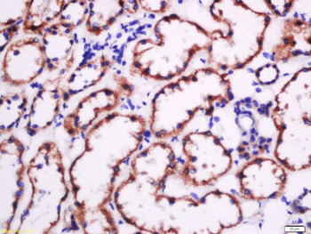
Immunohistochemistry of Anti-Fibronectin Antibody. Immunohistochemical assessment of proteins involved in blood coagulation in ob/ob pancreas. (E–J) Photomicrographs of representative pancreatic cryosections from lean control (E, F) and ob/ob (G, H) pancreas at 52 weeks of age labeled for Fibronectin (Green E, G) and von Willebrand Factor (Green, F, H) together with DAPI (blue). Areas enclosed by a broken line in (G, H) corresponds to (I, J) respectively. The areas in the lesions positive for Fibronectin and von Willebrand factor are not associated with any nucleated cells. Abbreviations; vWF, von Willebrand Factor; Exo, Exocrine tissue. Scale bar in (J) is 92 µm in (E–H) and 50 µm in (I, J).
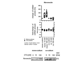
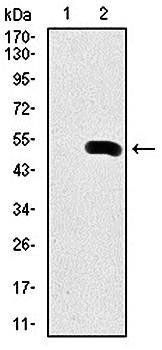
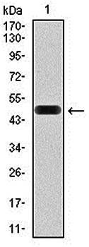
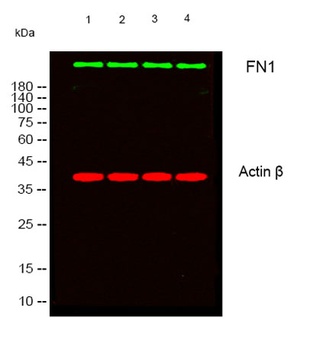
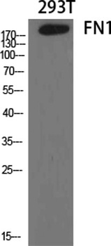
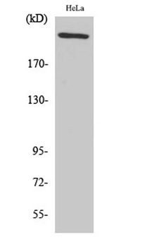
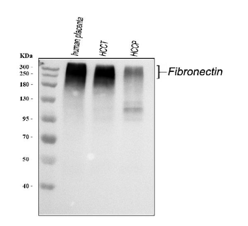
Western Blot of Anti-Fibronectin Antibody. Impact of OTA on cellular protein and mRNA amount in fibroblasts [NRK-49F]. OTA effect on fibronectin protein amount and mRNA abundance in NRK-49F under mono and co-culture conditions. Representative Western blots of proteins isolated from cells exposed to OTA. * indicates significant difference compared with the control group. Exposure of fibroblasts in monoculture to 10 or 100 nM OTA caused an increase of fibronectin protein amount.
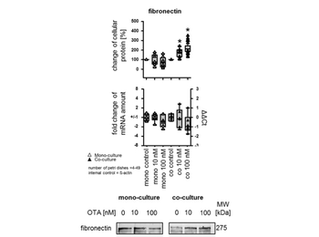
Western Blot of Anti-Fibronectin Antibody. Impact of OTA on the cellular protein and mRNA amount in renal epithelial cells [NRK-52E]. OTA effect on cellular fibronectin protein amount and mRNA abundance in NRK-52E under mono- and co-culture conditions. Representative Western blots of proteins isolated from cells exposed to OTA. * indicates significant difference compared with the control group. In the presence of fibroblasts, exposure to 10 nM OTA led to an increase of fibronectin protein amount. Incubation with 100 nM OTA led to an increase of fibronectin protein amount.
Quick Database Links
UniProt Details
− No UniProt data available
NCBI Reference Sequences
−Associated Accession Numbers
Curated reference sequences for the gene transcript and protein product| RefSeq | AAA53376.1 |
|---|
Documents Download
Datasheet
Product Information
Request a Document
Protocol Information
WB
Western Blot (IB, immunoblot)
IHC
Immunohistochemistry
ELISA
Enzyme-linked Immunosorbent Assay (EIA)
IP
Immunoprecipitation
FN1 Antibody (orb345363)

- 0.0
Based on 0 reviews
Participating in our Biorbyt product reviews program enables you to support fellow scientists by sharing your firsthand experience with our products.
Login to Submit a ReviewAvailable Sizes
Select a size below
Free Secondary Antibody (20 ul)0/0
Please add an antibody product to your cart first.