You have no items in your shopping cart.
ERBB2 Antibody
SKU: orb1926765
Description
Research Area
Signal Transduction
Images & Validation
−Item 1 of 2
| Tested Applications | IHC-P, WB |
|---|---|
| Dilution Range | WB - 1:1000, IHC-P - 1:1000 |
| Reactivity | Human |
Key Properties
−| Antibody Type | Primary Antibody |
|---|---|
| Host | Mouse |
| Clonality | Monoclonal |
| Isotype | IgG2b,κ |
| Molecular Weight | 137910 Da |
| Conjugation | Unconjugated |
Storage & Handling
−| Storage | Maintain refrigerated at 2-8°C for up to 2 weeks. For long term storage store at -20°C in small aliquots to prevent freeze-thaw cycles |
|---|---|
| Form/Appearance | Purified monoclonal antibody supplied in PBS with 0.09% (W/V) sodium azide. This antibody is purified through a protein G column, followed by dialysis against PBS. |
| Expiration Date | 12 months from date of receipt. |
| Disclaimer | For research use only |
Alternative Names
−HER2, MLN19, NEU, NGL
Similar Products
−- Item 1 of 6
ERBB2 Antibody [orb1929061]
FC, IF, IHC-P, WB
Human, Mouse, Rat
Rabbit
Polyclonal
Unconjugated
100 μl, 50 μl - Item 1 of 5
ERBB2/HER2/CD340 Antibody [orb1806342]
ELISA, FA, FACS, In vivo
Human, Monkey, Mouse
Monoclonal
Unconjugated
100 μg, 1 mg - Item 1 of 5
ERBB2/HER2/CD340 Antibody [orb1806343]
ELISA, FA, FACS, In vivo
Human, Mouse
Monoclonal
Unconjugated
100 μg, 1 mg - Item 1 of 1
Human Epidermal Growth Factor Receptor 2 (EGFR2) ELISA Kit [orb776097]
Human
0.63-40 ng/mL
0.223 ng/mL
96 T, 48 T - Item 1 of 1
Mouse Epidermal Growth Factor Receptor 2 (EGFR2) ELISA Kit [orb776523]
Mouse
0.32-20 ng/mL
0.121 ng/mL
48 T, 96 T


Quality Guarantee
Explore bioreagents carefree to elevate your research. All our products are rigorously tested for performance. If a product does not perform as described on its datasheet, our scientific support team will provide expert troubleshooting, a prompt replacement, or a refund. For full details, please see our Terms & Conditions and Buying Guide. Contact us at [email protected].
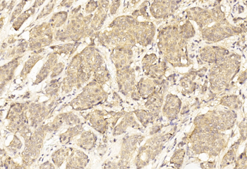
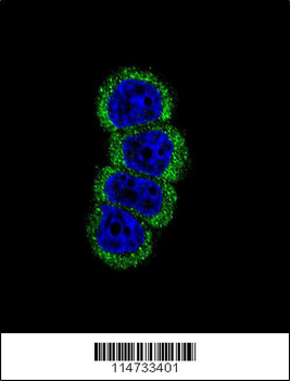
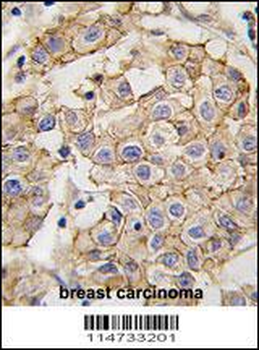
Immunohistochemical analysis of paraffin-embedded Human Breast cancer section using Pink1. Diluted at 1:1000 dilution. A undiluted biotinylated goat polyvalent antibody was used as the secondary, followed by DAB staining.
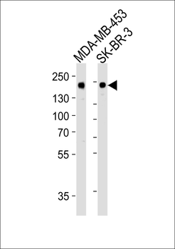
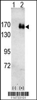
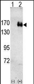
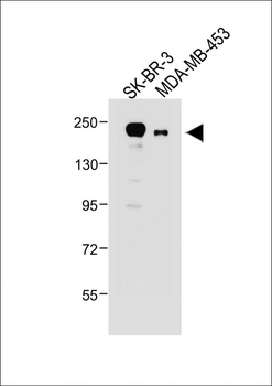
Western blot analysis of lysate from MDA-MB-453, SK-BR-3 cell line (from left to right), using ERBB2 Antibody. Diluted at 1:1000. A goat anti-mouse IgG H&L (HRP) at 1:10000 dilution was used as the secondary antibody. Lysate at 20 μg per lane.
Quick Database Links
UniProt
UniProt Details
− No UniProt data available
Documents Download
Datasheet
Product Information
Request a Document
Protocol Information
WB
Western Blot (IB, immunoblot)
IHC-P
Immunohistochemistry Paraffin
ERBB2 Antibody (orb1926765)

- 0.0
Based on 0 reviews
Participating in our Biorbyt product reviews program enables you to support fellow scientists by sharing your firsthand experience with our products.
Login to Submit a ReviewAvailable Sizes
Select a size below