You have no items in your shopping cart.
Description
Research Area
Immunology & Inflammation
Images & Validation
−Item 1 of 2
| Tested Applications | IHC-P, WB |
|---|---|
| Dilution Range | WB - 1:8000, IHC-P-Leica - 1:1000 |
| Reactivity | Human |
Key Properties
−| Host | Mouse |
|---|---|
| Clonality | Monoclonal |
| Isotype | IgG1 |
| Clone No. | B685EV47X3X3 |
| Immunogen | This EPCAM antibody is generated from mice immunized with a KLH conjugated synthetic peptide between 59-86 amino acids from human EPCAM. Antigen Region: 59-86 aa. |
| Target | EPCAM |
| Molecular Weight | 34932 Da |
| Conjugation | Unconjugated |
Storage & Handling
−| Storage | Maintain refrigerated at 2-8°C for up to 2 weeks. For long term storage store at -20°C in small aliquots to prevent freeze-thaw cycles |
|---|---|
| Form/Appearance | Purified monoclonal antibody supplied in PBS with 0.09% (W/V) sodium azide. This antibody is purified through a protein G column, followed by dialysis against PBS. |
| Expiration Date | 12 months from date of receipt. |
| Disclaimer | For research use only |
Alternative Names
−Epithelial cell adhesion molecule, Ep-CAM, Adenocarcinoma-associated antigen, Cell surface glycoprotein Trop-1, Epithelial cell surface antigen, Epithelial glycoprotein, EGP, Epithelial glycoprotein 314, EGP314, hEGP314, KS 1/4 antigen, KSA, Major gastrointestinal tumor-associated protein GA733-2, Tumor-associated calcium signal transducer 1, CD326, EPCAM, GA733-2, M1S2, M4S1, MIC18, TACSTD1, TROP1
Similar Products
−- Item 1 of 20
EpCAM Rabbit Polyclonal Antibody [orb10183]
FC, IF, IHC-Fr, IHC-P, WB
Porcine
Human, Mouse, Rat
Rabbit
Polyclonal
Unconjugated
50 μl, 100 μl, 200 μl - Item 1 of 11
EpCAM Antibody / Extracellular domain [orb606357]
FACS, IF, IHC-P, WB
Canine, Feline, Human
Mouse
Monoclonal
Unconjugated
100 μg, 20 μg - Item 1 of 11
EpCAM Antibody / Extracellular domain [orb2640454]
FACS, IF, IHC-P, WB
Canine, Feline, Human
Mouse
Monoclonal
Unconjugated
100 μg - Item 1 of 9
EpCAM Rabbit Polyclonal Antibody [orb1294311]
FC, IF, IHC, WB
Human, Mouse
Rabbit
Polyclonal
Unconjugated
100 μl, 25 μl - Item 1 of 5


Quality Guarantee
Explore bioreagents carefree to elevate your research. All our products are rigorously tested for performance. If a product does not perform as described on its datasheet, our scientific support team will provide expert troubleshooting, a prompt replacement, or a refund. For full details, please see our Terms & Conditions and Buying Guide. Contact us at [email protected].

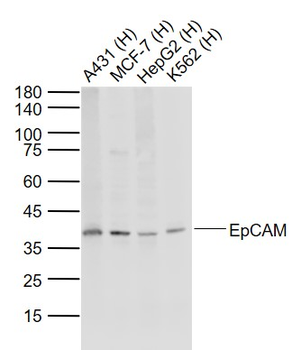
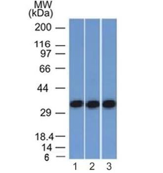
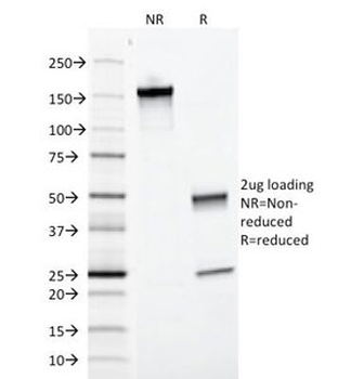
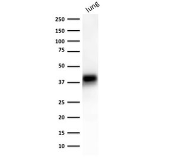
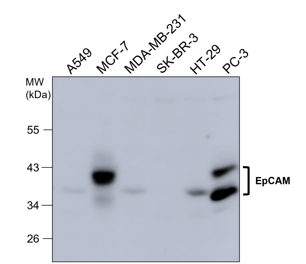
All lanes: Anti-EPCAM Antibody (N-term) at 1:8000 dilution. Lane 1: HT-29 whole cell lysate. Lane 2: MCF-7 whole cell lysate. Lysates/proteins at 20 µg per lane. Secondary Goat Anti-mouse IgG, (H+L), Peroxidase conjugated at 1/10000 dilution. Predicted band size: 39 kDa. Blocking/Dilution buffer: 5% NFDM/TBST.

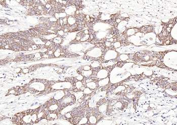
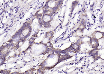
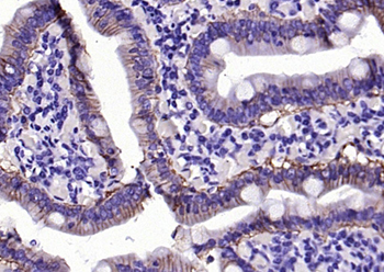
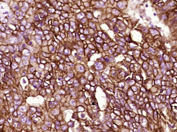
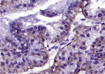
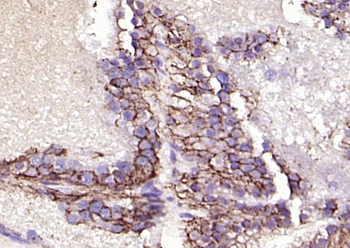
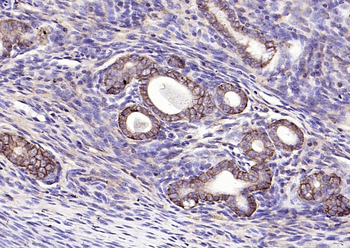
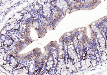
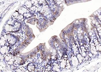
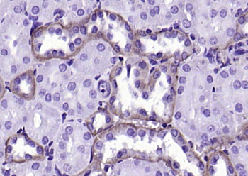
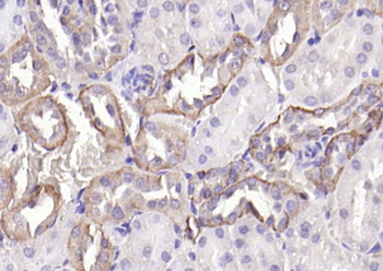
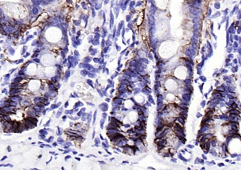
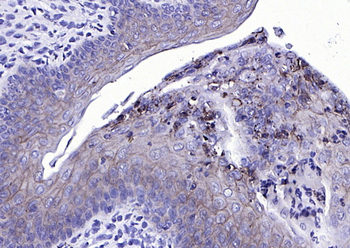
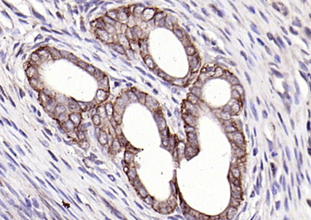
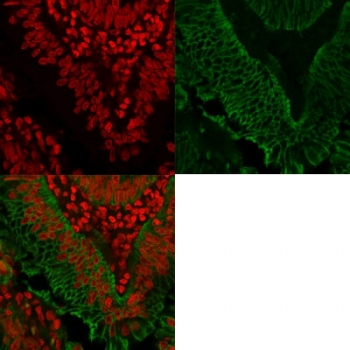
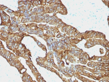
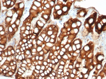
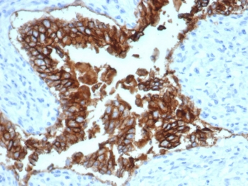
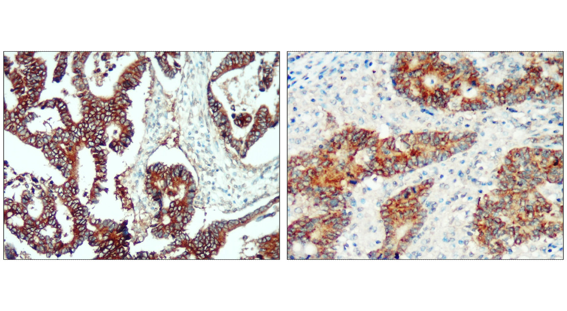
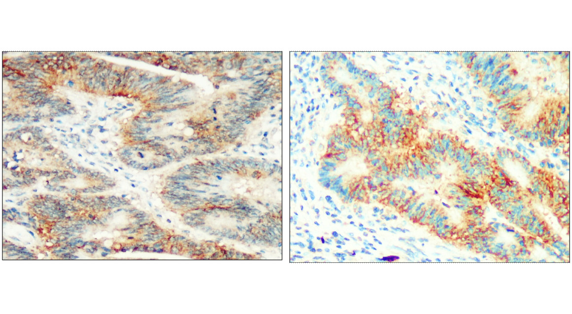
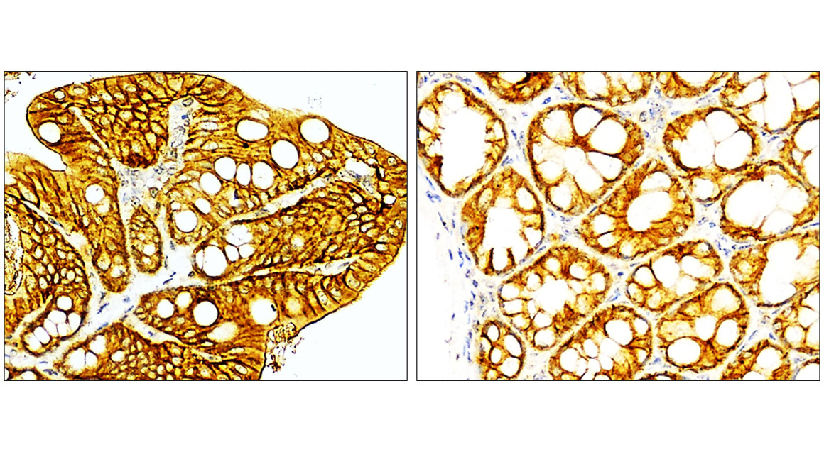
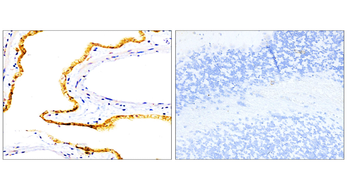
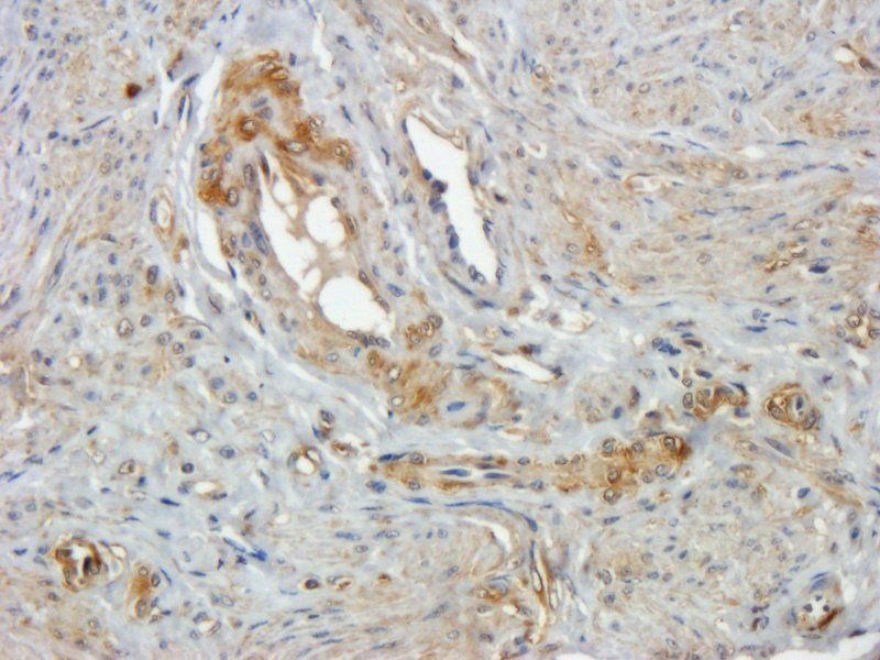
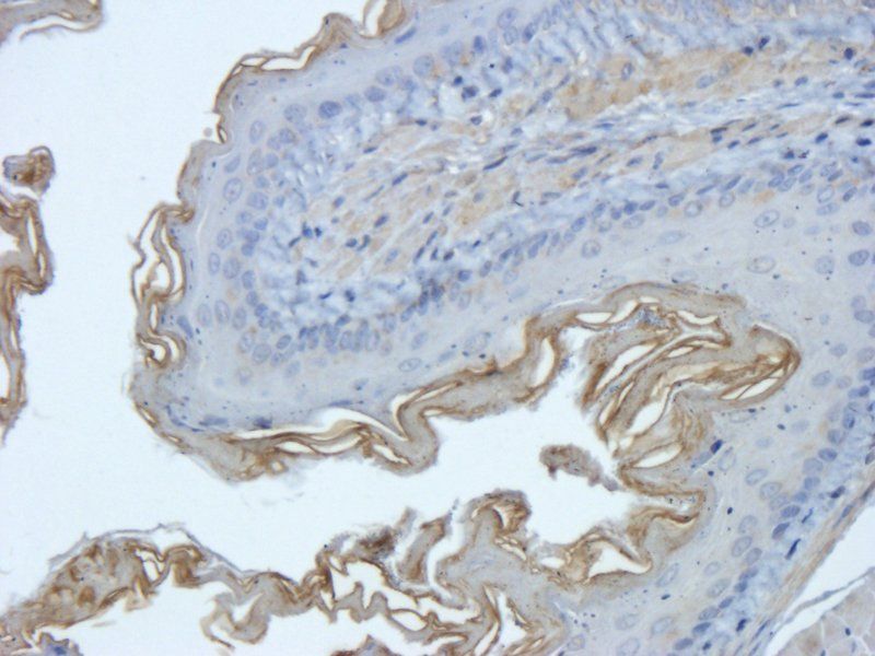
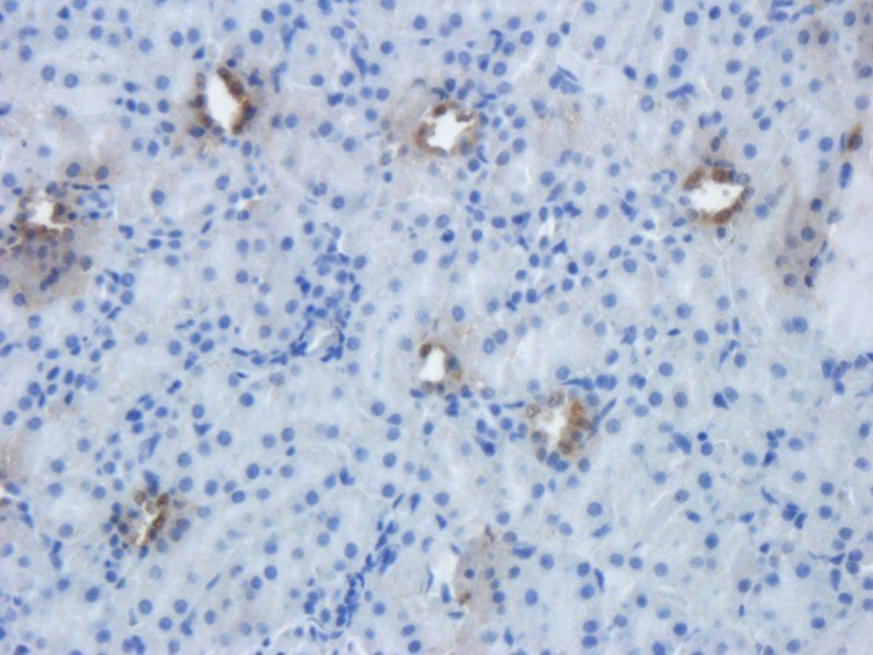
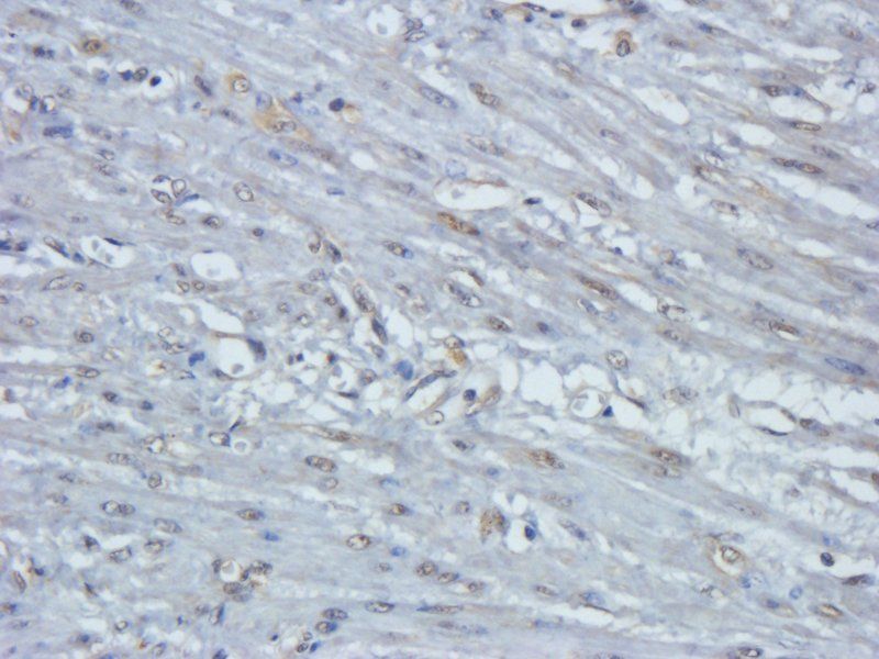
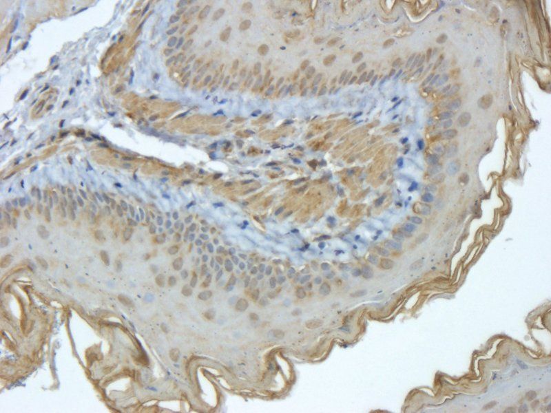
Immunohistochemical analysis of paraffin-embedded human appendix tissue performed on the Leica BOND RXm. Tissue was fixed with formaldehyde at room temperature; antigen retrieval was by heat mediation with a EDTA buffer (pH9.0). Samples were incubated with primary antibody (1:1000) for 1 hours at room temperature. A undiluted biotinylated CRF Anti-Polyvalent HRP Polymer antibody was used as the secondary Antibody.
Quick Database Links
UniProt Details
− No UniProt data available
NCBI Reference Sequences
−Associated Accession Numbers
Curated reference sequences for the gene transcript and protein product| Protein | NP_002345.2 |
|---|
Documents Download
Datasheet
Product Information
Request a Document
Protocol Information
WB
Western Blot (IB, immunoblot)
IHC-P
Immunohistochemistry Paraffin
EPCAM Antibody (orb1939252)

- 0.0
Based on 0 reviews
Participating in our Biorbyt product reviews program enables you to support fellow scientists by sharing your firsthand experience with our products.
Login to Submit a ReviewAvailable Sizes
Select a size below
Choose Conjugation or Carrier Free Version
Free Secondary Antibody (20 ul)0/0
Please add an antibody product to your cart first.