You have no items in your shopping cart.
Featured
Description
Images & Validation
−Item 1 of 12
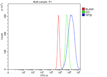
| Tested Applications | ICC, IF, IHC-P, WB |
|---|---|
| Dilution Range | IF/ICC: 1:50-400, IHC-P: 1:50-300, WB: 1:200-2000 |
| Reactivity | Guinea pig, Human, Mouse, Porcine, Rat |
Key Properties
−| Host | Rabbit |
|---|---|
| Clonality | Polyclonal |
| Isotype | IgG |
| Immunogen | KLH conjugated synthetic peptide derived from human ECP. Please contact us for the exact immunogen sequence. The peptide is available as orb12940. |
| Target | ECP |
| Molecular Weight | 18 kDa |
| Purity | Polyclonal antibodies are purified by peptide affinity chromatography |
| Conjugation | Unconjugated |
Storage & Handling
−| Storage | Maintain refrigerated at 2-8°C for up to 2 weeks. For long term storage store at -20°C in small aliquots to prevent freeze-thaw cycles. |
|---|---|
| Form/Appearance | 10 mM PBS, 0.02% sodium azide |
| Concentration | -100 μg (in 200 μl): 0.5 mg/ml-200 μg (in 400 μl): 0.5 mg/ml |
| Expiration Date | 12 months from date of receipt. |
| Disclaimer | For research use only |
Alternative Names
−anti-Cytotoxic ribonuclease antibody, anti-ECP antibody, anti-ECP_HUMAN antibody, anti-Eosinophil cationic protein antibody, anti-OTTHUMP00000164017 antibody, anti-Ribonuclease 3 antibody, anti-Ribonuclease, RNase A family, anti-RNase 3 antibody, anti-RNASE3 antibody, anti-RNS3 antibody
Similar Products
−- Item 1 of 10
TIP49A/RUVBL1 Rabbit Polyclonal Antibody [orb1173474]
ELISA, FC, ICC, IF, IHC, IP, WB
Human, Monkey, Mouse, Rat
Rabbit
Polyclonal
Unconjugated
100 μg - Item 1 of 3
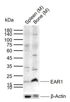
EAR1 Rabbit Polyclonal Antibody [orb13385]
FC, IF, IHC-Fr, IHC-P, WB
Human, Rat
Human, Mouse, Rat
Rabbit
Polyclonal
Unconjugated
50 μl, 100 μl, 200 μl - Item 1 of 5
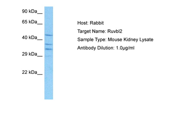
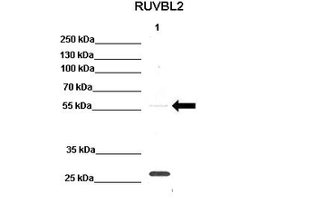
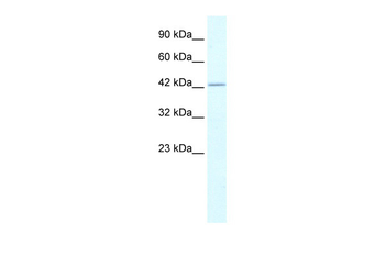
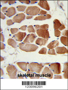
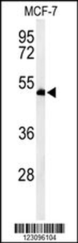
RUVBL2 Rabbit Polyclonal Antibody [orb574170]
IHC, WB
Bovine, Canine, Equine, Guinea pig, Rabbit, Rat, Zebrafish
Human, Mouse
Rabbit
Polyclonal
Unconjugated
100 μl - Item 1 of 4
- Item 1 of 4
Ribonuclease 3/RNASE3 Rabbit Polyclonal Antibody [orb443126]
ELISA, IHC, WB
Human, Mouse, Rat
Rabbit
Polyclonal
Unconjugated
100 μg

Quality Guarantee
Explore bioreagents carefree to elevate your research. All our products are rigorously tested for performance. If a product does not perform as described on its datasheet, our scientific support team will provide expert troubleshooting, a prompt replacement, or a refund. For full details, please see our Terms & Conditions and Buying Guide. Contact us at [email protected].

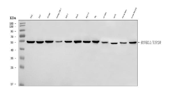
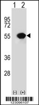
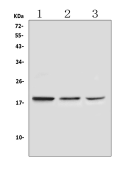
Western blot analysis of rat lymph node (lane 1), mouse colon (lane 2), rat spleen (lane 3), rat lung (lane 4), mouse uterus (lane 5), Hela (lane 6) using ECP antibody (1 ug/ml)
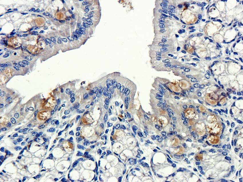
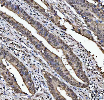
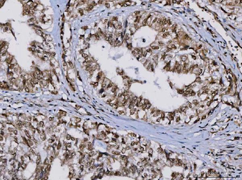
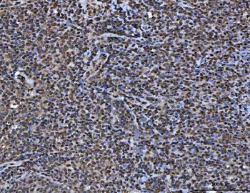
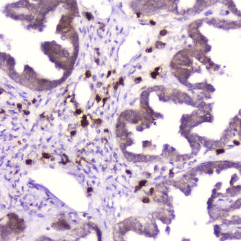
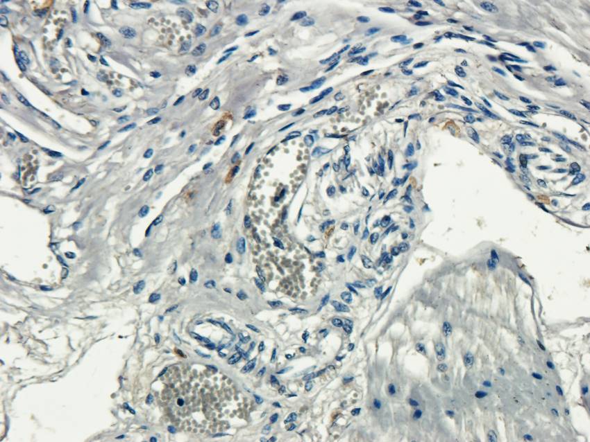
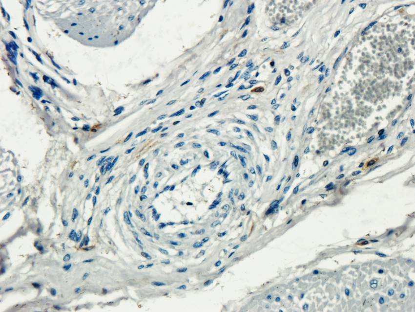
Immunohistochemical staining of paraffin embedded guinea pig rectum tissue using ECP antibody (primary antibody at 2.5 ug/ml)
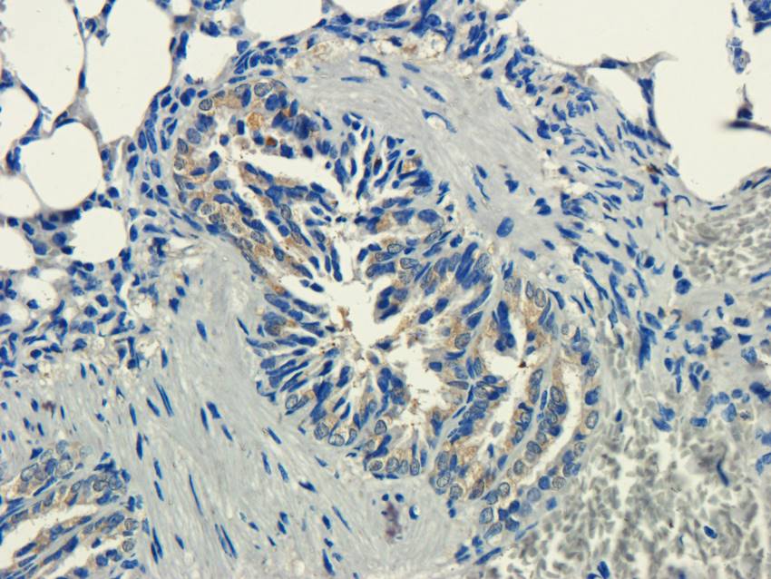
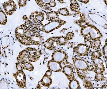
Immunohistochemical staining of guinea pig lung tissue using ECP antibody (dilution of primary antibody - 2.5 ug/ml)
IHC-P image of guinea pig rectum tissue using anti-ECP (dilution of primary antibody at 2.5 ug/ml)
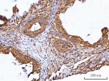
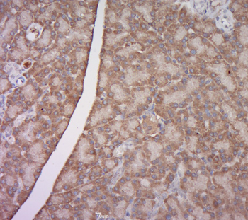
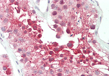
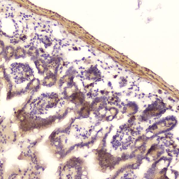
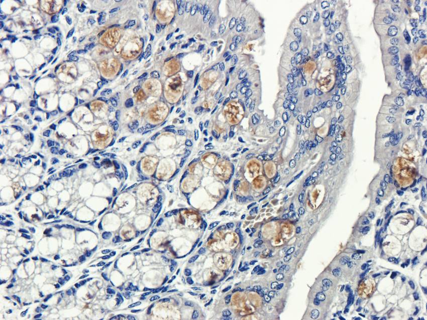
Immunohistochemical staining of pig uterus tissue using anti-ECP (dilution of primary antibody - 2.5 ug/ml)
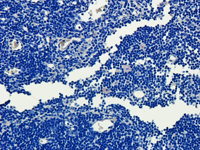
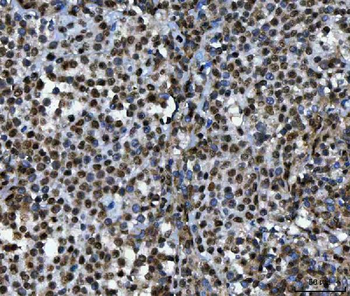
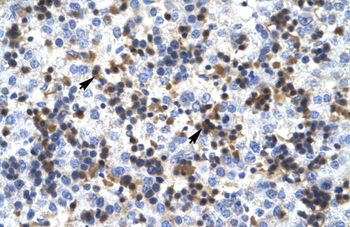
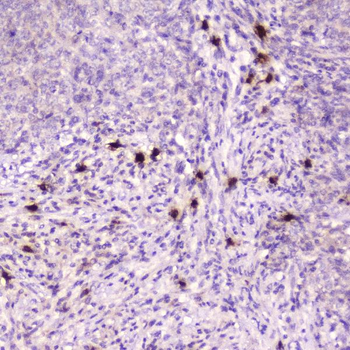
Immunohistochemical staining of rat lymph node tissue using ECP antibody (dilution of primary antibody - 2.5 ug/ml)
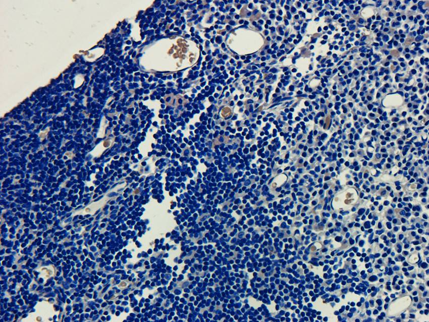
Immunohistochemical staining of paraffin embedded rat lymph node tissue using anti-ECP (primary antibody at 2.5 ug/ml)
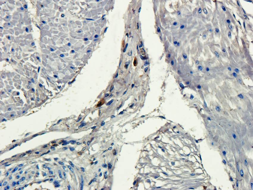
IHC-P staining of pig uterus tissue using anti-ECP (dilution at 2.5 ug/ml)
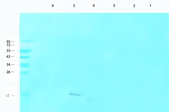
WB analysis of rat lymph node (lane 1), mouse colon (lane 2), rat spleen (lane 3), rat lung (lane 4), mouse uterus (lane 5), Hela (lane 6) using ECP antibody (1 ug/ml)
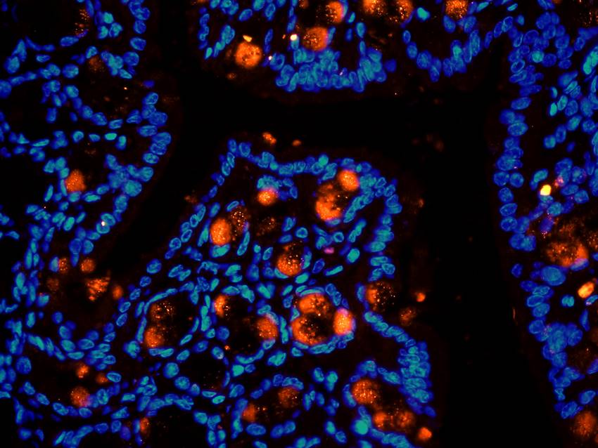
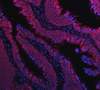
IF image of guinea pig rectum tissue using anti-ECP (primary antibody at 2.5 ug/ml)
Immunohistochemical staining of pig uterus tissue using anti-ECP (dilution of primary antibody - 2.5 ug/ml)
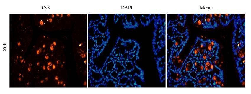
IF analysis of guinea pig rectum tissue using anti-ECP (dilution of primary antibody at 2.5 ug/ml)
Documents Download
Datasheet
Product Information
Request a Document
Protocol Information
WB
Western Blot (IB, immunoblot)
IHC-P
Immunohistochemistry Paraffin
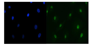
IF
Immunofluorescence
ICC
Immunocytochemistry
Xu X., Yu T., Dong L. et Eosinophils promote pulmonary matrix destruction and emphysema via Cathepsin L STTT, 8, 390 (2023)
ECP Rabbit Polyclonal Antibody (orb156688)

- 0.0
Based on 0 reviews
Participating in our Biorbyt product reviews program enables you to support fellow scientists by sharing your firsthand experience with our products.
Login to Submit a Review