You have no items in your shopping cart.
Dog1 antibody
SKU: orb2276582
Description
Images & Validation
−Item 1 of 3
| Tested Applications | IHC |
|---|---|
| Dilution Range | 1:100-1:200 |
| Reactivity | Human |
| Application Notes |
Key Properties
−| Host | Mouse |
|---|---|
| Clonality | Monoclonal |
| Isotype | IgG2b, kappa |
| Clone No. | MSVA-201M |
| Immunogen | Recombinant human DOG-1 protein fragment (aa 2-101) (exact sequence is proprietary) |
| Conjugation | Unconjugated |
Storage & Handling
−| Storage | Maintain refrigerated at 2-8°C for up to 2 weeks. For long term storage store at -20°C in small aliquots to prevent freeze-thaw cycles. |
|---|---|
| Expiration Date | 12 months from date of receipt. |
| Disclaimer | For research use only |
Alternative Names
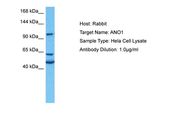
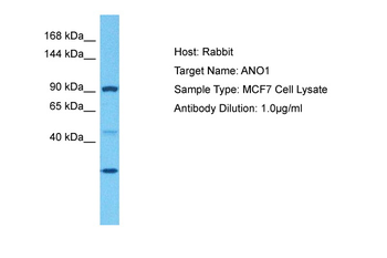
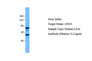
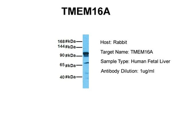
−Anoctamin 1; Calcium Activated Chloride Channel; Discovered On Gastrointestinal Stromal Tumors Protein 1; TAOS2; ORAOV2; TMEM16A
Similar Products
−- Item 1 of 7
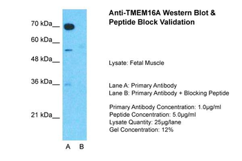
TMEM16A Rabbit Polyclonal Antibody [orb578503]
WB
Bovine, Canine, Equine, Guinea pig, Mouse, Rabbit, Rat
Human
Rabbit
Polyclonal
Unconjugated
100 μl - Item 1 of 3
- Item 1 of 3
- Item 1 of 3
- Item 1 of 3
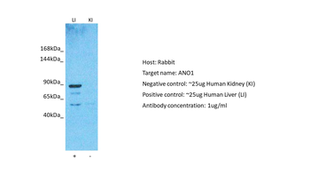
TMEM16A/ANO1 Rabbit Polyclonal Antibody [orb443147]
FC, IHC, WB
Human
Rabbit
Polyclonal
Unconjugated
100 μg


Quality Guarantee
Explore bioreagents carefree to elevate your research. All our products are rigorously tested for performance. If a product does not perform as described on its datasheet, our scientific support team will provide expert troubleshooting, a prompt replacement, or a refund. For full details, please see our Terms & Conditions and Buying Guide. Contact us at [email protected].

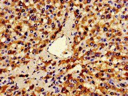
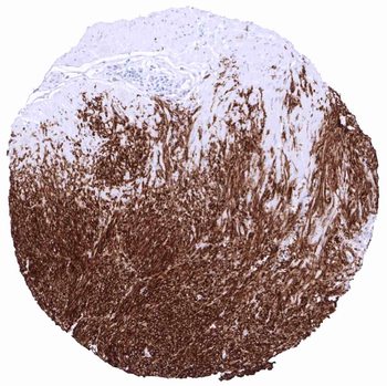
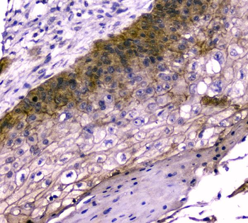
Dog1 Tumor periphery of a GIST showing intensive DOG1 staining in all tumor cells.

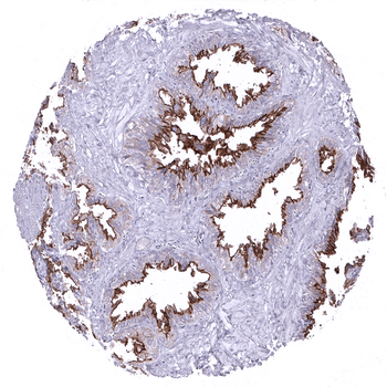
Parotid gland: Strong DOG1 staining of the apical membranes of secreting cells.

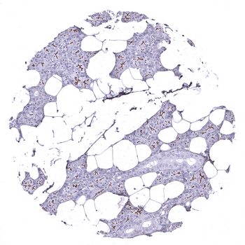
Seminal vesicle: Strong DOG1 staining of apical membranes of glandular cells.
Documents Download
Datasheet
Product Information
Request a Document
Dog1 antibody (orb2276582)

- 0.0
Based on 0 reviews
Participating in our Biorbyt product reviews program enables you to support fellow scientists by sharing your firsthand experience with our products.
Login to Submit a ReviewAvailable Sizes
Select a size below