You have no items in your shopping cart.
DAPI Staining Kit
SKU: orb219885
Description
Images & Validation
−Item 1 of 1
Key Properties
−Storage & Handling
−| Storage | Store at -20°C for one year. |
|---|---|
| Expiration Date | 12 months from date of receipt. |
| Disclaimer | For research use only |
Similar Products
−
DAPI solution (Nuclear Labeling) [orb1086014]
Unconjugated

Quality Guarantee
Explore bioreagents carefree to elevate your research. All our products are rigorously tested for performance. If a product does not perform as described on its datasheet, our scientific support team will provide expert troubleshooting, a prompt replacement, or a refund. For full details, please see our Terms & Conditions and Buying Guide. Contact us at [email protected].
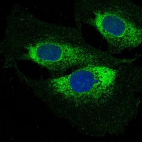
Immunofluorescent analysis staining in HeLa cells. Formalin-fixed cells were permeabilized with 0.1% Triton X-100 in TBS for 5-10 minutes and blocked with 3% BSA-PBS for 30 minutes at room temperature. Cells were probed with the primary antibody in 3% BSA-PBS and incubated overnight at 4 °C in a humidified chamber. Cells were washed with PBST and incubated with a FITC-conjugated secondary antibody (green) in PBS at room temperature in the dark. DAPI was used to stain the cell nuclei (blue).
Protocol Information
IF
Immunofluorescence
ICC
Immunocytochemistry
Filter by Applications
Filter by Species
M El Assar, B García-Gómez, JM La Fuente, M Alonso-Isa, JI Martínez-Salamanca Targeting TRPC-5 Channel Inhibition to Improve Penile Vascular Function in Erectile Dysfunction International Journal of Molecular Sciences, (2025)
Huanli Zhao 1, Xuejun Wu 2, Shumeng Yang 3, Lili Jiang 4, Huiying Yu 3, Yubin L Formononetin Alleviates the Inflammatory Response Induced by Carotid Balloon Injury in Rats via the PP2A/MAPK Axis Immunol Invest, (2025)
Applications
IF
Reactivity
Rat
Fábio Trindade 1, João Almeida-Coelho 1, Cláudia Sousa-Mendes 1, Francisca Saraiva 1, Maria L Arbonés 2 3, Adelino Leite-Moreira 1 4, Rui Vitorino # 1 5 6, Inês Falcão-Pires Myocardial phosphoproteomics unveils a key role of DYRK1A in aortic valve replacement-induced reverse remodelling Basic Res Cardiol, (2025)
Applications
IF
Reactivity
Human
Zahra Kohan 1, Jalal Shayegh 2, Tohid Kazemi 3, Ehsan Ahmadpour 4, Shabnam Babaei 5, Najibeh Shekari 5 Immune modulation by melanoma-derived exosomes: suppression of BALB/c mice splenic cell proliferation, induction of apoptosis, and cell cycle arrest Mol Biol Rep ., (2025)
Zhang, Min et al. Neuroprotective effects of miR-30c on rats with cerebral ischemia/reperfusion injury by targeting SOX9 Pathol Res Pract, 216, 153271 (2020)
DAPI Staining Kit (orb219885)

- 0.0
Based on 0 reviews
Participating in our Biorbyt product reviews program enables you to support fellow scientists by sharing your firsthand experience with our products.
Login to Submit a ReviewAvailable Sizes
Select a size below