You have no items in your shopping cart.
Featured
Description
Research Area
Epigenetics & Chromatin
Images & Validation
−Item 1 of 10
| Tested Applications | ICC, IF, IHC-Fr, IHC-P, WB |
|---|---|
| Dilution Range | WB=1:500-2000, IHC-P=1:100-500, IHC-F=1:100-500, ICC/IF=1:100-500, IF=1:100-500 |
| Reactivity | Human, Mouse, Rat |
| Predicted Reactivity | Bovine |
Related Conjugates & Formulations
−Key Properties
−| Antibody Type | Primary Antibody |
|---|---|
| Host | Rabbit |
| Clonality | Polyclonal |
| Isotype | IgG |
| Immunogen | KLH conjugated synthetic peptide derived from human CDK7 (1-80/346aa) |
| Target | CDK7 |
| Molecular Weight | 40 kDa |
| Purification | Affinity purified by Protein A |
| Conjugation | Unconjugated |
Storage & Handling
−| Storage | Maintain refrigerated at 2-8°C for up to 2 weeks. For long term storage store at -20°C in small aliquots to prevent freeze-thaw cycles. |
|---|---|
| Form/Appearance | Liquid |
| Buffer/Preservatives | 0.01M TBS (pH7.4) with 1% rAlbumin, 0.02% Proclin300 and 50% Glycerol. |
| Concentration | 1mg/ml |
| Expiration Date | 12 months from date of receipt. |
| Disclaimer | For research use only |
Alternative Names
−CAK; CAK1; CDKN7; HCAK; MO15; STK1; p39MO15; CDK7_HUMAN; CDK7; 39 kDa protein kinase (p39 Mo15); CDK-activating kinase 1; Cell division protein kinase 7; Serine/threonine-protein kinase 1; TFIIH basal transcription factor complex kinase subunit; 2.7.11.22; 2.7.11.23; cyclin dependent kinase 7; cyclin-dependent kinase 7 (homolog of Xenopus MO15 cdk-activating kinase); cyclin-dependent kinase 7 (MO15 homolog, Xenopus laevis, cdk-activating kinase)
Similar Products
−- Item 1 of 6
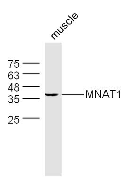
MNAT1 Rabbit Polyclonal Antibody [orb312140]
FC, ICC, IF, IHC, WB
Human, Mouse, Rat
Rabbit
Polyclonal
Unconjugated
100 μg - Item 1 of 4
Phospho-CDK7(T170) Antibody [orb1931280]
DOT, IHC-P, WB
Human, Mouse
Rabbit
Polyclonal
Unconjugated
50 μl, 100 μl - Item 1 of 4
Phospho-CDK7 (Thr170) Rabbit Polyclonal Antibody [orb312194]
ICC, IF, IHC-Fr, IHC-P, WB
Canine, Equine, Mouse, Porcine, Rabbit, Rat
Human
Rabbit
Polyclonal
Unconjugated
50 μl, 100 μl, 200 μl - Item 1 of 3
MNAT1 Rabbit Polyclonal Antibody [orb101218]
FC, IF, IHC-Fr, IHC-P, WB
Bovine, Canine, Equine, Gallus, Human, Porcine, Rabbit, Sheep
Mouse, Rat
Rabbit
Polyclonal
Unconjugated
50 μl, 100 μl, 200 μl - Item 1 of 3

Quality Guarantee
Explore bioreagents carefree to elevate your research. All our products are rigorously tested for performance. If a product does not perform as described on its datasheet, our scientific support team will provide expert troubleshooting, a prompt replacement, or a refund. For full details, please see our Terms & Conditions and Buying Guide. Contact us at [email protected].
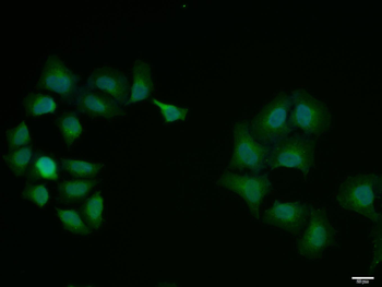
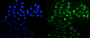
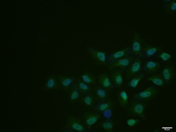
Hela cell, 4% Paraformaldehyde-fixed, Triton X-100 at room temperature for 20 min, Blocking buffer (normal goat serum) at 37°C for 20 min, Antibody incubation with (CDK7) polyclonal Antibody, Unconjugated (orb10361) 1:100, 90 minutes at 37°C, followed by a conjugated Goat Anti-Rabbit IgG antibody at 37°C for 90 minutes, DAPI (blue) was used to stain the cell nuclei.
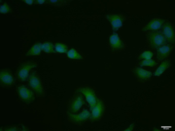
Hela cell, 4% Paraformaldehyde-fixed, Triton X-100 at room temperature for 20 min, Blocking buffer (normal goat serum) at 37°C for 20 min, Antibody incubation with (CDK7) polyclonal Antibody, Unconjugated (orb10361) 1:100, 90 minutes at 37°C, followed by a conjugated Goat Anti-Rabbit IgG antibody at 37°C for 90 minutes, DAPI (blue) was used to stain the cell nuclei.
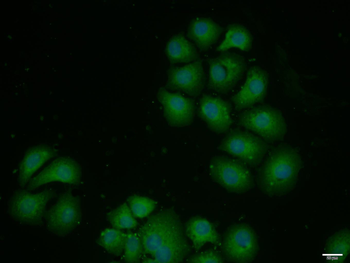
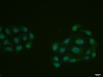
HepG2 cell, 4% Paraformaldehyde-fixed, Triton X-100 at room temperature for 20 min, Blocking buffer (normal goat serum) at 37°C for 20 min, Antibody incubation with (CDK7) polyclonal Antibody, Unconjugated (orb10361) 1:100, 90 minutes at 37°C, followed by a conjugated Goat Anti-Rabbit IgG antibody at 37°C for 90 minutes, DAPI (blue) was used to stain the cell nuclei.
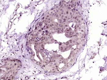
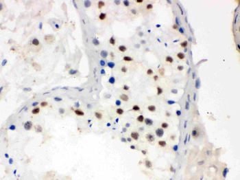
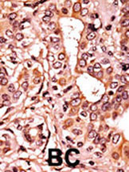
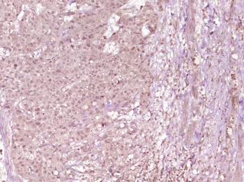
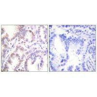
Paraformaldehyde-fixed, paraffin embedded (Human breast carcinoma), Antigen retrieval by boiling in sodium citrate buffer (pH6.0) for 15 min, Block endogenous peroxidase by 3% hydrogen peroxide for 20 minutes, Blocking buffer (normal goat serum) at 37°C for 30 min, Antibody incubation with (CDK7) Polyclonal Antibody, Unconjugated (orb10361) at 1:400 overnight at 4°C, followed by operating according to SP Kit (Rabbit) instructionsand DAB staining.
Paraformaldehyde-fixed, paraffin embedded (human cervical carcinoma), Antigen retrieval by boiling in sodium citrate buffer (pH6.0) for 15 min, Block endogenous peroxidase by 3% hydrogen peroxide for 20 minutes, Blocking buffer (normal goat serum) at 37°C for 30 min, Antibody incubation with (CDK7) Polyclonal Antibody, Unconjugated (orb10361) at 1:400 overnight at 4°C, followed by a conjugated secondary for 20 minutes and DAB staining.
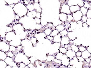
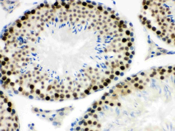
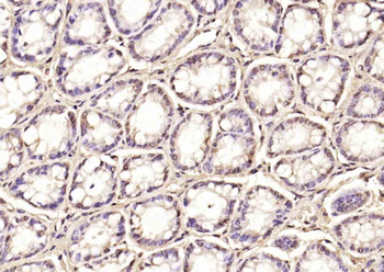
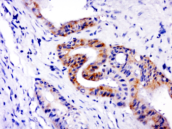
Paraformaldehyde-fixed, paraffin embedded (mouse lung), Antigen retrieval by boiling in sodium citrate buffer (pH6.0) for 15 min, Block endogenous peroxidase by 3% hydrogen peroxide for 20 minutes, Blocking buffer (normal goat serum) at 37°C for 30 min, Antibody incubation with (CDK7) Polyclonal Antibody, Unconjugated (orb10361) at 1:200 overnight at 4°C, followed by operating according to SP Kit (Rabbit) instructionsand DAB staining.
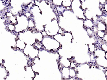
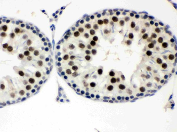
Paraformaldehyde-fixed, paraffin embedded (rat lung), Antigen retrieval by boiling in sodium citrate buffer (pH6.0) for 15 min, Block endogenous peroxidase by 3% hydrogen peroxide for 20 minutes, Blocking buffer (normal goat serum) at 37°C for 30 min, Antibody incubation with (CDK7) Polyclonal Antibody, Unconjugated (orb10361) at 1:200 overnight at 4°C, followed by operating according to SP Kit (Rabbit) instructionsand DAB staining.
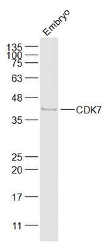
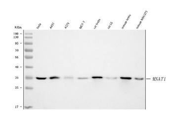
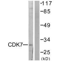
Sample: Embryo (Mouse) Lysate at 40 ug, Primary: Anti-CDK7 (orb10361) at 1/1000 dilution, Secondary: IRDye800CW Goat Anti-Rabbit IgG at 1/20000 dilution, Predicted band size: 40 kD, Observed band size: 40 kD.
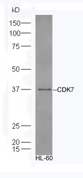
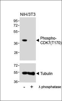
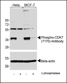
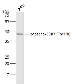
Sample: HL-60 Cell lysate, Primary:Anti-CDK7 (orb10361) at 1:300, Secondary: HRP conjugated Goat-Anti-rabbit IgG (orb572747) at 1:5000, Predicted band size: 40 kD, Observed band size: 37 kD.
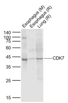
Sample: Lane 1: Esophagus (Mouse) Lysate at 40 ug, Lane 2: Esophagus (Rat) Lysate at 40 ug, Lane 3: Lung (Rat) Lysate at 40 ug, Primary: Anti-CDK7 (orb10361) at 1/1000 dilution, Secondary: IRDye800CW Goat Anti-Rabbit IgG at 1/20000 dilution, Predicted band size: 43 kD, Observed band size: 43 kD.
Quick Database Links
Gene Symbol
CDK7
UniProt
UniProt Details
− No UniProt data available
Documents Download
Datasheet
Product Information
Request a Document
Protocol Information
WB
Western Blot (IB, immunoblot)
IHC-P
Immunohistochemistry Paraffin
IHC-Fr
Immunohistochemistry Frozen
IF
Immunofluorescence
ICC
Immunocytochemistry
CDK7 Rabbit Polyclonal Antibody (orb10361)

- 0.0
Based on 0 reviews
Participating in our Biorbyt product reviews program enables you to support fellow scientists by sharing your firsthand experience with our products.
Login to Submit a ReviewAvailable Sizes
Select a size below
Free Secondary Antibody (20 ul)0/0
Please add an antibody product to your cart first.