You have no items in your shopping cart.
Description
Images & Validation
−Item 1 of 5
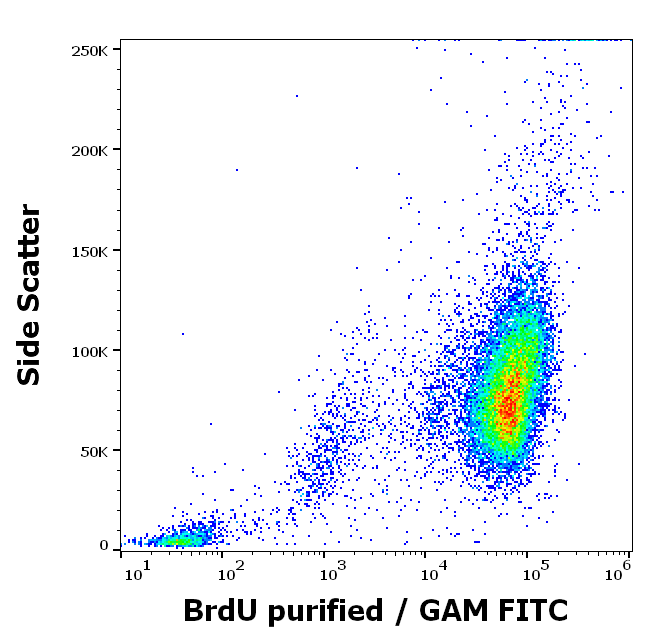
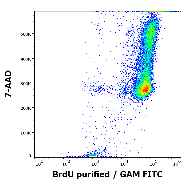
| Tested Applications | DOT, ELISA, FC, IF, IHC, WB |
|---|---|
| Dilution Range | ELISA: 1:2000 - 1:10000, FC: 1:50-1:100, IHC: 1:100-1:500, IF: 1:500-1:3000, WB: 1:2000 - 1:5000 |
| Reactivity | Other |
| Application Notes |
Key Properties
−| Antibody Type | Primary Antibody |
|---|---|
| Host | Mouse |
| Clonality | Monoclonal |
| Isotype | IgG |
| Clone No. | 29G6.E8 |
| Immunogen | Anti-BrdU monoclonal antibody was produced in mice by repeated immunizations prepared via immunizations with BromodeoxyUridine-KLH followed by hybridoma development. |
| Purity | Anti-BrdU Monoclonal Antibody was purified from ascites fluid by Protein A chromatography. This antibody reacts strongly with BrdU. Cross-reactivity is observed with CldU and IdU. |
| Conjugation | Unconjugated |
Storage & Handling
−| Storage | Store BrdU Antibody at -20° C prior to opening. Aliquot contents and freeze at -20° C or below for extended storage. Avoid cycles of freezing and thawing. Centrifuge product if not completely clear after standing at room temperature. This product is stable for several weeks at 4° C as an undiluted liquid. Dilute only prior to immediate use. |
|---|---|
| Form/Appearance | Liquid (sterile filtered) |
| Buffer/Preservatives | Preservative: 0.01% (w/v) Sodium Azide. Stabilizer: None; Buffer: 0.02 M Potassium Phosphate, 0.15 M Sodium Chloride, pH 7.2 |
| Concentration | 1.0 mg/ml |
| Expiration Date | 12 months from date of receipt. |
| Dry Ice Shipping | Please note: This product requires shipment on dry ice. A dry ice surcharge will apply. |
| Disclaimer | For research use only |
Alternative Names
−mouse anti-BrdU Antibody, Bromodeoxyuridine, 5-bromo-2'-deoxyuridine, BrdU
Similar Products
−- Item 1 of 6
Mouse Bromodeoxyuridine / BrdU Antibody [orb98204]
FC, ICC, IHC-Fr, IHC-P
Other
Mouse
Monoclonal
Unconjugated
0.1 mg - Item 1 of 3
BrdU (Proliferation Marker) Rabbit Polyclonal Antibody [orb10204]
ELISA
All
All
Rabbit
Polyclonal
Unconjugated
50 μl, 100 μl, 200 μl - Item 1 of 1
Mouse BrdU (bromodeoxyuridine), conjugated with FITC Antibody [orb108872]
FACS, IHC-P
Mouse
Monoclonal
FITC
100 μg - Item 1 of 1
- Item 1 of 3


Quality Guarantee
Explore bioreagents carefree to elevate your research. All our products are rigorously tested for performance. If a product does not perform as described on its datasheet, our scientific support team will provide expert troubleshooting, a prompt replacement, or a refund. For full details, please see our Terms & Conditions and Buying Guide. Contact us at [email protected].

Bromodeoxyuridine (BrdU) chemical structure representation.

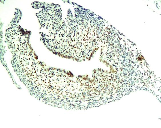
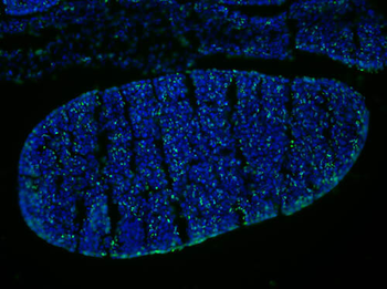
Immunofluorescence Microscopy of Mouse Anti-BrdU antibody. Tissue: OCT-embedded E10.5 mouse embryo. Localization: 20X, section through the developing hindbrain. Fixation: 4% PFA. Antigen retrieval: not required. Primary antibody: BrdU antibody at 1:1000 for overnight at 4°C in 0.4% PBS + Triton with 1% normal sheep serum. Secondary antibody: Alexa Fluor 488 Anti-Mouse secondary antibody at 1:200 for 45 min at RT. Staining: Double labeled (green/blue) cells represent cells that were actively dividing.
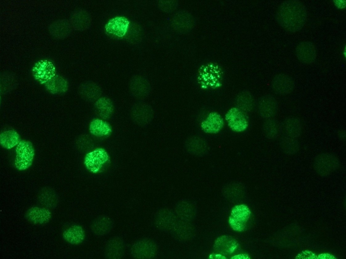
Immunofluorescence Microscopy of Mouse Anti-BrdU antibody. Tissue: OCT-embedded E10.5 mouse embryo. Localization: 20X, section through the developing limb bud. Fixation: 4% PFA. Antigen retrieval: not required. Primary antibody: BrdU antibody at 1:1000 in 0.4% PBS + Triton with 1% normal sheep serum overnight at 4°C. Secondary antibody: Alexa Fluor 488 Anti-Mouse secondary antibody at 1:200 for 45 min at RT. Staining: Double labeled (green/blue) cells represent cells that were actively dividing.
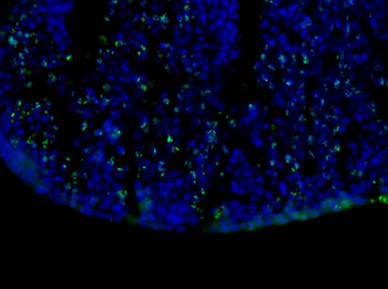
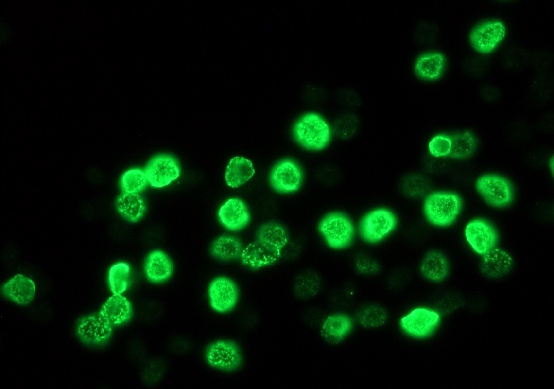
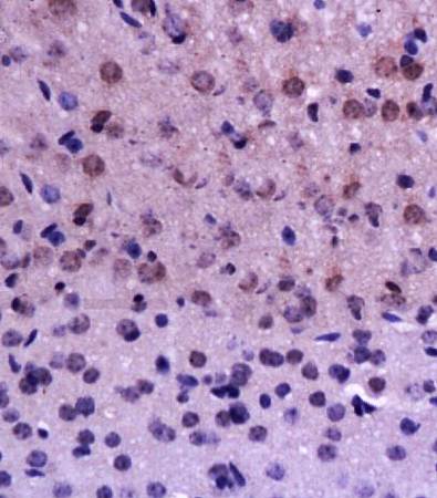
Immunofluorescence Microscopy of Mouse Anti-BrdU antibody. Tissue: OCT-embedded E10.5 mouse embryo. Localization: 40X, section through the developing limb bud. Fixation: 4% PFA. Antigen retrieval: not required. Primary antibody: BrdU antibody at 1:500 in 0.4% PBS + Triton with 1% normal sheep serum overnight at 4°C. Secondary antibody: Alexa Fluor 488 Anti-Mouse secondary antibody at 1:200 for 45 min at RT. Staining: Double labeled (green/blue) cells represent cells that were actively dividing.

Western blot of Anti-BrdU antibody. Lane 1: loading control. Lane 2: FdU. Lane 3: BrdU. Lane 4: IdU. Lane 5 CldU. Load: 20 µg per lane. Primary antibody: Anti-BrdU antibody at 1:1000 for overnight at 4°C. Secondary antibody: IRDye800™ mouse secondary antibody at 1:10000 for 45 min at RT. Block: 5% BLOTTO overnight at 4°C. Predicted: BrdU. Other band(s): cross reactive bands observed for other nucleoside analogs, IdU and CldU.
Documents Download
Datasheet
Product Information
Request a Document
Protocol Information
WB
Western Blot (IB, immunoblot)
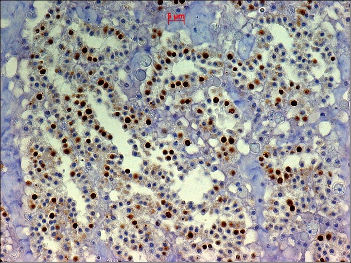
IHC
Immunohistochemistry
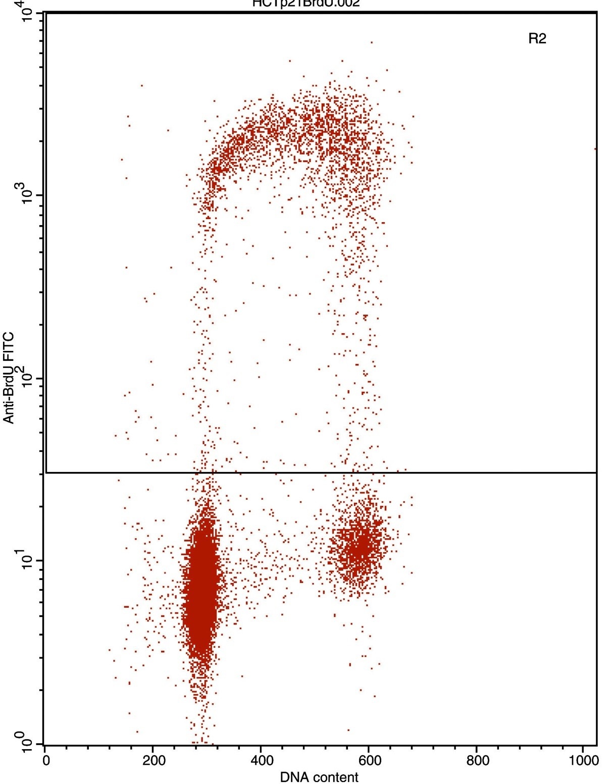
FC
Flow Cytometry
IF
Immunofluorescence
ELISA
Enzyme-linked Immunosorbent Assay (EIA)
DOT
Dot Blot
BrdU Antibody (orb344495)

- 0.0
Based on 0 reviews
Participating in our Biorbyt product reviews program enables you to support fellow scientists by sharing your firsthand experience with our products.
Login to Submit a ReviewAvailable Sizes
Select a size below
Choose Conjugation or Carrier Free Version
Free Secondary Antibody (20 ul)0/0
Please add an antibody product to your cart first.