You have no items in your shopping cart.
Description
Research Area
Cell Biology, Immunology & Inflammation, Signal Transduction, Stem Cell & Developmental Biology
Images & Validation
−Item 1 of 7
| Tested Applications | IHC, WB |
|---|---|
| Reactivity | Human |
| Predicted Reactivity | Bovine, Canine, Equine, Guinea pig, Mouse, Porcine, Rat, Yeast, Zebrafish |
Key Properties
−| Host | Rabbit |
|---|---|
| Clonality | Polyclonal |
| Immunogen | The immunogen is a synthetic peptide directed towards the N terminal region of human ATP6V0A2 |
| Target | ATP6V0A2 |
| Protein Sequence | Synthetic peptide located within the following region: INRADIPLPEGEASPPAPPLKQVLEMQEQLQKLEVELREVTKNKEKLRKN |
| Molecular Weight | 98kDa |
| Purification | Affinity Purified |
| Conjugation | Unconjugated |
Storage & Handling
−| Storage | Maintain refrigerated at 2-8°C for up to 2 weeks. For long term storage store at -20°C in small aliquots to prevent freeze-thaw cycles. |
|---|---|
| Buffer/Preservatives | Liquid. Purified antibody supplied in 1x PBS buffer with 0.09% (w/v) sodium azide and 2% sucrose. |
| Concentration | 0.5 mg/ml |
| Expiration Date | 12 months from date of receipt. |
| Disclaimer | For research use only |
Alternative Names
−A2, RTF, TJ6, WSS, a2V, ARCL, J6B7, STV1, TJ6M, TJ6S, VPH1, ARCL2A, ATP6A2, ATP6N1D
Similar Products
−- Item 1 of 2
ATP6V0A2 Rabbit Polyclonal Antibody [orb235031]
IF, WB
Human, Monkey, Mouse, Rat
Rabbit
Polyclonal
Unconjugated
30 μl, 100 μl, 200 μl, 50 μl - Item 1 of 2
- Item 1 of 1

ATP6V0A2 polyclonal antibody [orb645702]
WB
Human, Mouse
Rabbit
Polyclonal
Unconjugated
200 μl, 100 μl, 50 μlATP6V0A2 Rabbit Polyclonal Antibody [orb154891]
ELISA, IHC, WB
Mouse, Rat
Human
Rabbit
Polyclonal
Unconjugated
100 μg


Quality Guarantee
Explore bioreagents carefree to elevate your research. All our products are rigorously tested for performance. If a product does not perform as described on its datasheet, our scientific support team will provide expert troubleshooting, a prompt replacement, or a refund. For full details, please see our Terms & Conditions and Buying Guide. Contact us at [email protected].
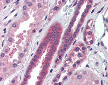
Anti-ATP6V0A2 antibody IHC staining of human kidney. Immunohistochemistry of formalin-fixed, paraffin-embedded tissue after heat-induced antigen retrieval. Antibody concentration 5 ug/ml.
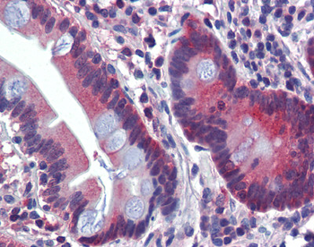
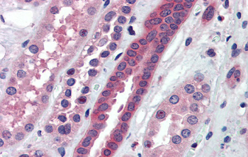
Anti-ATP6V0A2 antibody IHC staining of human small intestine. Immunohistochemistry of formalin-fixed, paraffin-embedded tissue after heat-induced antigen retrieval. Antibody concentration 5 ug/ml.
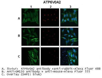
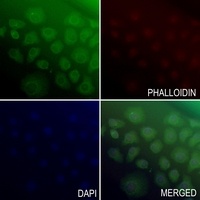
Application: IHC/Immunofluorescence, Species+tissue/cell type: A. untransfected HeLa cells, B. mATP6V0A2-FLAG transfected HeLa cells, C. mATP6V0A2 (partial) transfected HeLa cells, Primary antibody dilution: 1:250, Secondary antibody: Anti-rabbit AlexaFluor 488, Secondary antibody dilution: 1:1000.
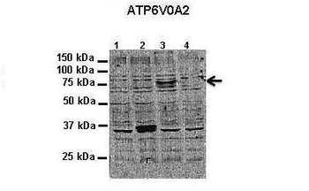
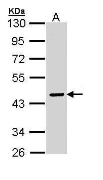
Application: Western blotting, Species+tissue/cell type: HeLa cells, How many ug'sof tissue/cell lysate run on the gel: 1. 10 ug untransfected HeLa lysate, 2. 10 ug mATP6V0A2 (Partial) transfected HeLa lysate, 3. 10 ug mATP6V0A2-FLAG transfected HeLa lysate, 4. 10 ug mATP6V0A1-FLAG transfected HeLa lysate, Primary antibody dilution: 1:300, Secondary antibody: Anti-rabbit-HRP, Secondary antibody dilution: 1:1000.
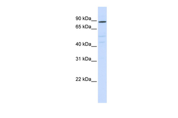
ATP6V0A2 antibody - N-terminal region (orb578447) validated by WB using HeLa cells at 1:300.
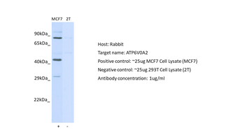
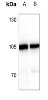
Positive control (+): MCF7 (N10), Negative control (-): HEK293 (HEK293), Antibody concentration: 1 ug/ml.
Immunohistochemistry with Human kidney lysate tissue at an antibody concentration of 5.0 ug/ml using anti-ATP6V0A2 antibody (orb578447).
Documents Download
Datasheet
Product Information
Request a Document
Protocol Information
WB
Western Blot (IB, immunoblot)
IHC
Immunohistochemistry
ATP6V0A2 Rabbit Polyclonal Antibody (orb578447)

- 0.0
Based on 0 reviews
Participating in our Biorbyt product reviews program enables you to support fellow scientists by sharing your firsthand experience with our products.
Login to Submit a ReviewAvailable Sizes
Select a size below
Free Secondary Antibody (20 ul)0/0
Please add an antibody product to your cart first.