You have no items in your shopping cart.
ATG16L Antibody
SKU: orb1933395
Description
Research Area
Cell Biology
Images & Validation
−Item 1 of 6
| Tested Applications | IF, IHC-P, WB |
|---|---|
| Dilution Range | WB - 1:1000, IHC-P - 1:100-500 |
| Reactivity | Human, Mouse |
Key Properties
−| Host | Rabbit |
|---|---|
| Clonality | Polyclonal |
| Isotype | Rabbit IgG |
| Molecular Weight | 68265 Da |
| Conjugation | Unconjugated |
Storage & Handling
−| Storage | Maintain refrigerated at 2-8°C for up to 2 weeks. For long term storage store at -20°C in small aliquots to prevent freeze-thaw cycles |
|---|---|
| Form/Appearance | Purified polyclonal antibody supplied in PBS with 0.09% (W/V) sodium azide. This antibody is prepared by Saturated Ammonium Sulfate (SAS) precipitation followed by dialysis against PBS. |
| Expiration Date | 12 months from date of receipt. |
| Disclaimer | For research use only |
Alternative Names
−APG16L
Similar Products
−- Item 1 of 4
ATG16L1/ATG16L Antibody [orb1537867]
ELISA, ICC, IF, IHC, IHC-P, WB
Human, Mouse, Rat
Rabbit
Polyclonal
Unconjugated
50 μg - Item 1 of 5
- Item 1 of 5
ATG16L1 Rabbit Polyclonal Antibody [orb1098121]
ELISA, ICC, IF, IHC, WB
Human
Rabbit
Polyclonal
Unconjugated
100 μg - Item 1 of 4
- Item 1 of 2


Quality Guarantee
Explore bioreagents carefree to elevate your research. All our products are rigorously tested for performance. If a product does not perform as described on its datasheet, our scientific support team will provide expert troubleshooting, a prompt replacement, or a refund. For full details, please see our Terms & Conditions and Buying Guide. Contact us at [email protected].
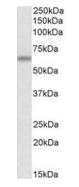
Western blot of APG16L (L92) Pab in mouse brain tissue lysate.
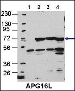
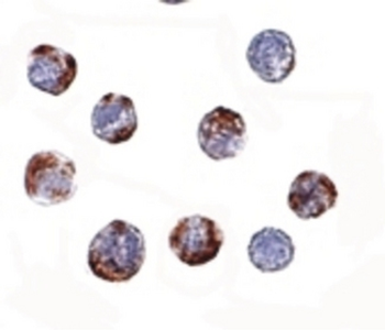
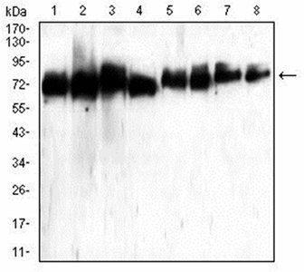
Cos7, HEK293, MEF, and Hela cells, left to right, respectively.
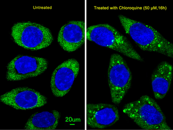
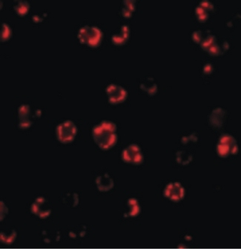
Immunofluorescent analysis of U251 cells, using ATG16L Antibody. U251 cells (right) were treated with Chloroquine (50 μM, 16 h). Diluted at 1:25 dilution. Alexa Fluor 488-conjugated goat anti-rabbit lgG at 1:400 dilution was used as the secondary antibody (green). DAPI was used to stain the cell nuclear (blue).
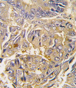
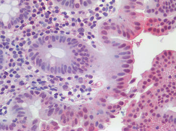
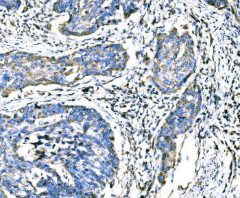
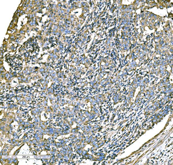
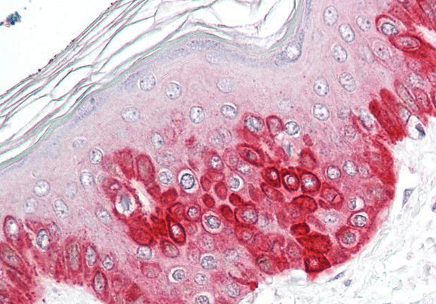
Formalin-fixed and paraffin-embedded human colon carcinoma tissue reacted with Autophagy APG16L antibody (L176), which was peroxidase-conjugated to the secondary antibody, followed by DAB staining. This data demonstrates the use of this antibody for immunohistochemistry; clinical relevance has not been evaluated.
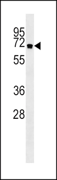
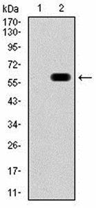
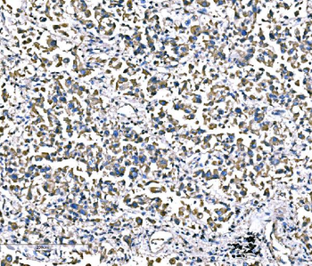
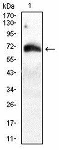
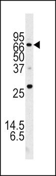
APG16L Antibody western blot analysis in NCI-H460 cell line lysates (35 ug/lane). This demonstrates the APG16L antibody detected the APG16L protein (arrow).
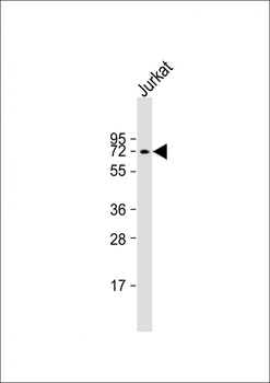
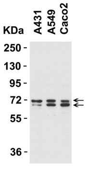
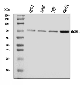
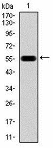
Anti-ATG16L Antibody at 1:1000 dilution + Jurkat whole cell lysate. Lysates/proteins at 20 µg per lane. Secondary Goat Anti-Rabbit IgG, (H+L), Peroxidase conjugated at 1/10000 dilution. Predicted band size: 68 kDa. Blocking/Dilution buffer: 5% NFDM/TBST.
Quick Database Links
UniProt
UniProt Details
− No UniProt data available
Documents Download
Datasheet
Product Information
Request a Document
Protocol Information
WB
Western Blot (IB, immunoblot)
IHC-P
Immunohistochemistry Paraffin
IF
Immunofluorescence
ATG16L Antibody (orb1933395)

- 0.0
Based on 0 reviews
Participating in our Biorbyt product reviews program enables you to support fellow scientists by sharing your firsthand experience with our products.
Login to Submit a ReviewAvailable Sizes
Select a size below