You have no items in your shopping cart.
Featured
KO/KD
Validated
Validated
Description
Research Area
Cell Biology
Images & Validation
−Item 1 of 8
| Tested Applications | ELISA, ICC, IHC-P, KO/KD Validated, WB |
|---|---|
| Reactivity | Human |
| Predicted Reactivity | Mouse, Rat |
Key Properties
−| Antibody Type | Primary Antibody |
|---|---|
| Host | Rabbit |
| Clonality | Polyclonal |
| Isotype | IgG |
| Immunogen | Anti-ASC antibody (orb1239217) was raised against a peptide corresponding to 14 amino acids near the carboxy terminus of human ASC. The immunogen is located within the last 50 amino acids of ASC. |
| Target | PYCARD |
| Molecular Weight | Predicted: 22kDObserved: 22 kD |
| Purification | ASC Antibody is affinity chromatography purified via peptide column. |
| Conjugation | Unconjugated |
Storage & Handling
−| Storage | Maintain refrigerated at 2-8°C for up to 2 weeks. For long term storage store at -20°C in small aliquots to prevent freeze-thaw cycles. |
|---|---|
| Form/Appearance | Liquid |
| Buffer/Preservatives | ASC Antibody is supplied in PBS containing 0.02% sodium azide. |
| Concentration | 1 mg/mL |
| Expiration Date | 12 months from date of receipt. |
| Disclaimer | For research use only |
Alternative Names
−ASC Antibody: ASC, TMS, TMS1, CARD5, TMS-1, ASC, Apoptosis-associated speck-like protein containing a CARD, Caspase recruitment domain-containing protein 5, hASC
Similar Products
−- Item 1 of 12
ASC/TMS1/PYCARD Rabbit Polyclonal Antibody [orb1728091]
ELISA, FC, ICC, IF, IHC, WB
Human
Rabbit
Polyclonal
Unconjugated
100 μg - Item 1 of 7
ASC/TMS1 Rabbit Polyclonal Antibody [orb100371]
WB
Human
Rabbit
Polyclonal
Unconjugated
50 μl, 100 μl, 200 μl - Item 1 of 8
- Item 1 of 7
- Item 1 of 4
Pycard (rodent) Antibody [orb107673]
ELISA, IF
Bovine, Canine, Porcine
Human, Mouse, Rat
Polyclonal
Unconjugated
100 μg


Quality Guarantee
Explore bioreagents carefree to elevate your research. All our products are rigorously tested for performance. If a product does not perform as described on its datasheet, our scientific support team will provide expert troubleshooting, a prompt replacement, or a refund. For full details, please see our Terms & Conditions and Buying Guide. Contact us at [email protected].
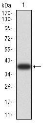
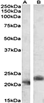
Western Blot Validation in Human HL60 Cells. Loading: 15 µg of lysates per lane. Antibodies: ASC orb1239217, (1 µg/mL) in the absence (A) or presence (B) of blocking peptide, 1h incubation at RT in 5% NFDM/TBST. Secondary: Goat anti-rabbit IgG HRP conjugate at 1:10000 dilution.
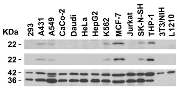
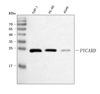
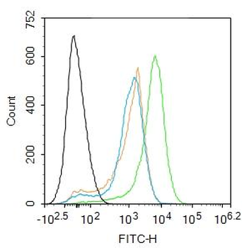
Independent Antibody Validation (IAV) via Protein Expression Profile in Cell Lines. Loading: 15 µg of lysates per lane. Antibodies: ASC orb1239217, (2 µg/mL), ASC, (2 µg/mL), beta-actin (1 µg/mL) and GAPDH (0.02 µg/mL), 1h incubation at RT in 5% NFDM/TBST. Secondary: Goat anti-rabbit or goat anti-mouse (for ASC 39001) IgG HRP conjugate at 1:10000 dilution.
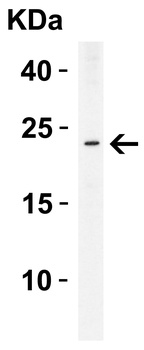
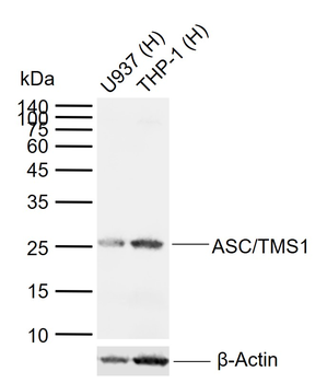
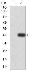
Western Blot Validation in Human THP-1 Cells. Loading: 15 µg of lysate per lane. Antibodies: ASC orb1239217, (2 µg/mL), 1h incubation at RT in 5% NFDM/TBST. Secondary: Goat anti-rabbit IgG HRP conjugate at 1:10000 dilution.
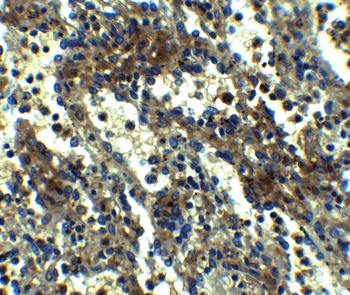
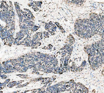
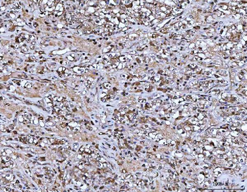
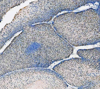
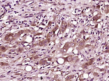
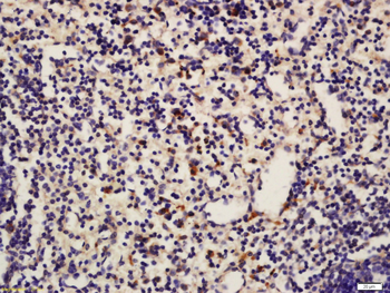
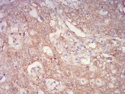
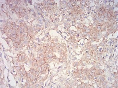
Immunohistochemistry Validation of ASC in Human Spleen Tissue. Immunohistochemical analysis of paraffin-embedded human spleen tissue using anti-ASC antibody (orb1239217) at 2.5 µg/ml. Tissue was fixed with formaldehyde and blocked with 10% serum for 1 h at RT; antigen retrieval was by heat mediation with a citrate buffer (pH6). Samples were incubated with primary antibody overnight at 4°C. A goat anti-rabbit IgG H&L (HRP) at 1/250 was used as secondary. Counter stained with Hematoxylin.
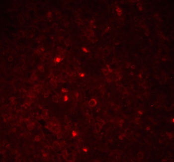
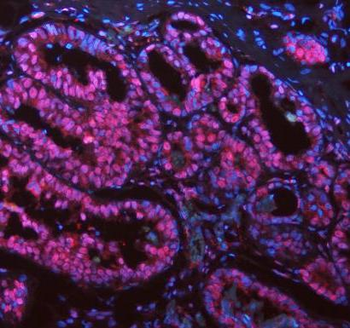
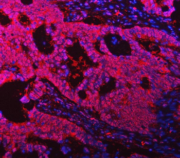
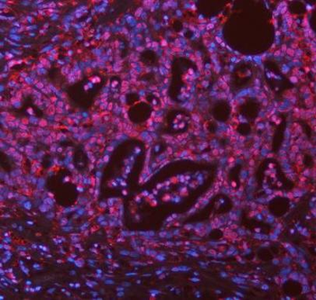
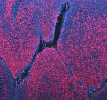
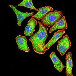
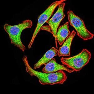
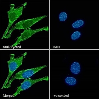
Immunofluorescence Validation of ASC in Human Spleen Tissue. Immunofluorescent analysis of 4% paraformaldehyde-fixed Human Spleen Tissue labeling ASC with orb1239217 at 20 µg/mL, followed by goat anti-rabbit IgG secondary antibody at 1/500 dilution (red).
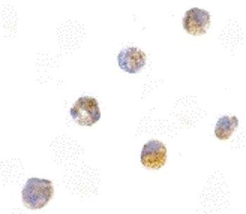
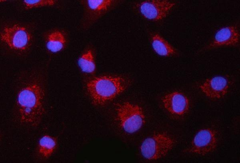
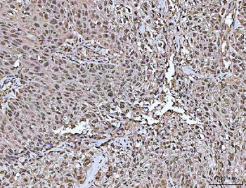
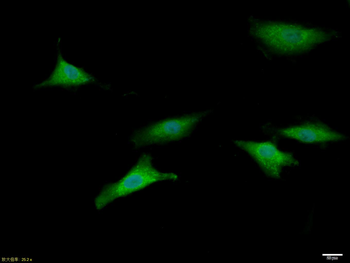
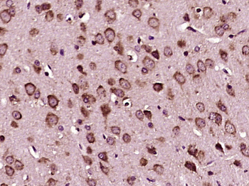
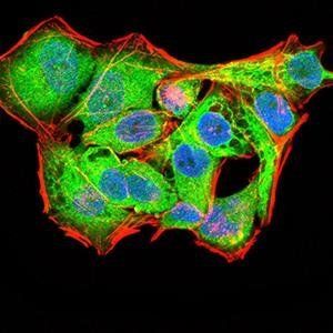
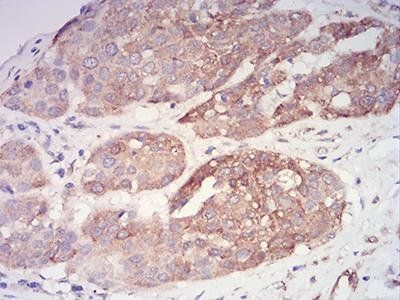
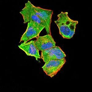
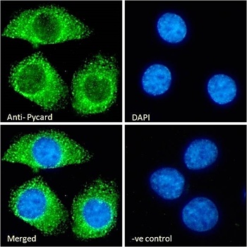
Immunocytochemistry Validation of ASC in HL60 Cells. Immunocytochemical analysis of HL60 cells using anti-ASC antibody (orb1239217) at 5 µg/ml. Cells was fixed with formaldehyde and blocked with 10% serum for 1 h at RT; antigen retrieval was by heat mediation with a citrate buffer (pH6). Samples were incubated with primary antibody overnight at 4°C. A goat anti-rabbit IgG H&L (HRP) at 1/250 was used as secondary. Counter stained with Hematoxylin.
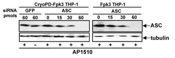
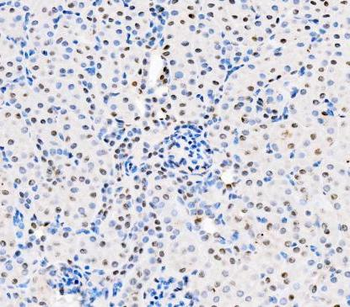
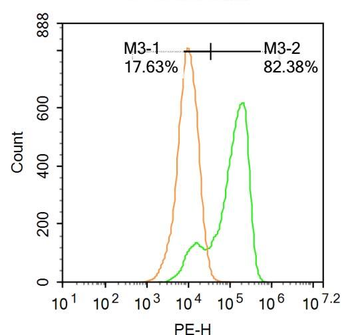
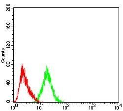
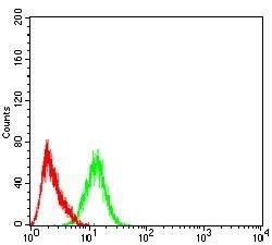
KD Validation of ASC in THP-1 Cells (Dowds et al., 2004). Immunofluorescence analysis with anti-ASC antibodies (orb1239217) was performed for BIM in 293 cells transfected with GFP siRNA or ASC siRNA. ASC expression was disrupted after ASC siRNA knockdown.
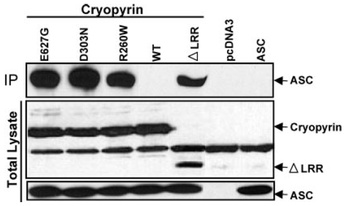
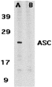
Overexpression Validation of ASC in HEK293T Cells (Dowds et al., 2004). Western blot analysis with anti-ASC antibodies (orb1239217) was performed for ASC in HEK293T cells transfected with pcDNA3-ASC.
Documents Download
Datasheet
Product Information
Request a Document
Protocol Information
WB
Western Blot (IB, immunoblot)
IHC-P
Immunohistochemistry Paraffin
ICC
Immunocytochemistry
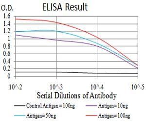
ELISA
Enzyme-linked Immunosorbent Assay (EIA)
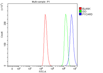
PYCARD Antibody (orb1239217)

- 0.0
Based on 0 reviews
Participating in our Biorbyt product reviews program enables you to support fellow scientists by sharing your firsthand experience with our products.
Login to Submit a ReviewAvailable Sizes
Select a size below
Free Secondary Antibody (20 ul)0/0
Please add an antibody product to your cart first.