You have no items in your shopping cart.
Description
Images & Validation
−Item 1 of 2
| Tested Applications | ELISA, IF, IHC, WB |
|---|---|
| Dilution Range | ELISA: 1:4,000 - 1:16,000, IHC: 20-40 µg/ml, WB: 1:500 - 1:2,000 |
| Reactivity | Human, Mouse |
| Application Notes |
Key Properties
−| Antibody Type | Primary Antibody |
|---|---|
| Host | Rabbit |
| Clonality | Polyclonal |
| Isotype | IgG |
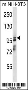
| Immunogen | This affinity purified antibody was prepared from whole rabbit serum produced by repeated immunizations with a synthetic peptide corresponding to an internal region near amino acids 775-800 of mouse ASAP1 protein. |
| Target | Asap1 |
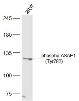
| Purity | This affinity-purified antibody is directed against the phosphorylated form of mouse ASAP1 protein at the pY782 residue. The product was affinity purified from monospecific antiserum by immunoaffinity purification. Antiserum was first purified against the phosphorylated form of the immunizing peptide. The resultant affinity purified antibody was then cross-adsorbed against the non-phosphorylated form of the immunizing peptide. Reactivity occurs against mouse ASAP1 pY782 protein and the antibody is specific for the phosphorylated form of the protein. Reactivity with non-phosphorylated mouse ASAP1 is minimal by ELISA. A BLAST analysis was used to suggest cross reactivity with ASAP1 proteins from human, chicken, bovine, dog, rat and chimpanzee based on 100% homology with the immunizing sequence. Reactivity against homologues from other sources is not known. |
| Conjugation | Unconjugated |
Storage & Handling
−| Storage | Store vial at -20° C prior to opening. Aliquot contents and freeze at -20° C or below for extended storage. Avoid cycles of freezing and thawing. Centrifuge product if not completely clear after standing at room temperature. This product is stable for several weeks at 4° C as an undiluted liquid. Dilute only prior to immediate use. |
|---|---|
| Form/Appearance | Liquid (sterile filtered) |
| Buffer/Preservatives | Preservative: 0.01% (w/v) Sodium Azide. Stabilizer: None; Buffer: 0.02 M Potassium Phosphate, 0.15 M Sodium Chloride, pH 7.2 |
| Concentration | 1.00 mg/mL |
| Expiration Date | 12 months from date of receipt. |
| Dry Ice Shipping | Please note: This product requires shipment on dry ice. A dry ice surcharge will apply. |
| Disclaimer | For research use only |
Alternative Names
−rabbit anti-ASAP1 pY782 Antibody, ASAP-1, ASAP 1, Development and differentiation enhancing factor 1 antibody, 130 kDa phosphatidylinositol 4 5 biphosphate dependent ARF1 GTPase activating protein antibody, ADP ribosylation factor directed GTPase activating protein 1 antibody, AMAP 1 antibody
Similar Products
−- Item 1 of 1
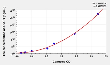
Human ADP-Ribosylation Factor GTPase Activating Protein 1 (ASAP1) ELISA Kit [orb1736790]
Human
0.32-20 ng/mL
0.18 ng/mL
48 T, 96 T - Item 1 of 1
- Item 1 of 1
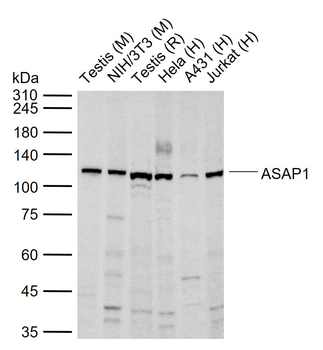
ASAP1 Rabbit Polyclonal Antibody [orb4397]
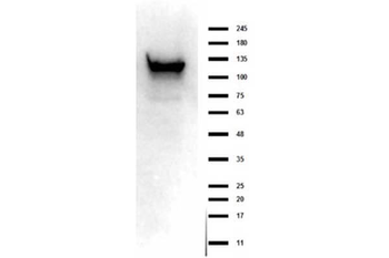
WB
Bovine, Canine, Equine, Rabbit
Human, Mouse, Rat
Rabbit
Polyclonal
Unconjugated
50 μl, 100 μl, 200 μl - Item 1 of 1
Phospho-ASAP1 (Tyr782) Rabbit Polyclonal Antibody [orb4398]
WB
Bovine, Canine, Equine, Gallus, Guinea pig, Mouse, Porcine, Rat
Human
Rabbit
Polyclonal
Unconjugated
50 μl, 100 μl, 200 μl - Item 1 of 1
Quality Guarantee
Explore bioreagents carefree to elevate your research. All our products are rigorously tested for performance. If a product does not perform as described on its datasheet, our scientific support team will provide expert troubleshooting, a prompt replacement, or a refund. For full details, please see our Terms & Conditions and Buying Guide. Contact us at [email protected].
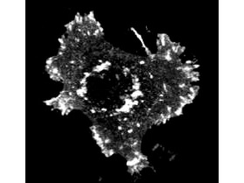
Immunofluorescent microscopy using Biorbyt's Affinity Purified anti-ASAP1 pY782 antibody shows detection of phosphorylated ASAP1 present in mouse NIH3T3 cells transfected with activated Src. Specific staining is not present when antibody is pre-incubated with the immunizing peptide prior to reaction with cells.
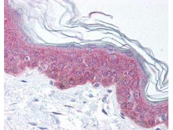
Biorbyt's affinity purified anti-ASAP1 pY782 antibody was used at 20 µg/ml to detect signal in a variety of tissues including multi-human, multi-brain and multi-cancer slides. This image shows moderate intracellular positive staining in epidermal keratinocytes in human skin at 40X. Tissue was formalin-fixed and paraffin embedded. The image shows localization of the antibody as the precipitated red signal, with a hematoxylin purple nuclear counterstain.
Documents Download
Datasheet
Product Information
Request a Document
Protocol Information
WB
Western Blot (IB, immunoblot)
IHC
Immunohistochemistry
IF
Immunofluorescence
ELISA
Enzyme-linked Immunosorbent Assay (EIA)
Ruggiero, Carmen et al. The KDEL receptor signalling cascade targets focal adhesion kinase on focal adhesions and invadopodia Oncotarget, 9, 10228-10246 (2018)
Asap1 Antibody (orb345529)

- 0.0
Based on 0 reviews
Participating in our Biorbyt product reviews program enables you to support fellow scientists by sharing your firsthand experience with our products.
Login to Submit a ReviewAvailable Sizes
Select a size below
Choose Conjugation or Carrier Free Version
Free Secondary Antibody (20 ul)0/0
Please add an antibody product to your cart first.