You have no items in your shopping cart.
Description
Research Area
Cancer Biology
Images & Validation
−Item 1 of 2
| Tested Applications | ELISA, FC, ICC, IHC-P, IP, WB |
|---|---|
| Reactivity | Human, Primate |
| Application Notes |
Key Properties
−| Clonality | Monoclonal |
|---|---|
| Isotype | Mouse IgG2a |
| Clone No. | BP53-12 |
| Immunogen | Bacterially expressed full-length wild-type p53 |
| Target | p53 |
| Purification | Purified antibody is conjugated with biotin LC-NHS ester under optimum conditions and unconjugated antibody and free biotin are removed by size-exclusion chromatography. |
| Conjugation | Biotin |
Storage & Handling
−| Storage | Store at 2-8°C. Do not freeze. |
|---|---|
| Buffer/Preservatives | Phosphate buffered saline (PBS), pH 7.4, 15 mM sodium azide |
| Concentration | 1 mg/ml |
| Expiration Date | 12 months from date of receipt. |
| Disclaimer | For research use only |
Alternative Names
−BCC7, TRP53, TP53, LFS1
Similar Products
−- Item 1 of 1
Human Proline dehydrogenase 1, Mitochondrial (PRODH) ELISA Kit [orb1736755]
Human
78.13-5000 pg/mL
33.8 pg/mL
96 T, 48 T - Item 1 of 1
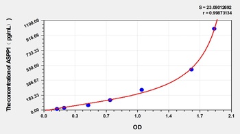
Human Apoptosis-stimulating of p53 Protein 1 (ASPP1) ELISA Kit [orb1173549]
Human
15.63-1000 pg/mL
6.6 pg/mL
96 T, 48 T - Item 1 of 3

- Item 1 of 2

Quality Guarantee
Explore bioreagents carefree to elevate your research. All our products are rigorously tested for performance. If a product does not perform as described on its datasheet, our scientific support team will provide expert troubleshooting, a prompt replacement, or a refund. For full details, please see our Terms & Conditions and Buying Guide. Contact us at [email protected].
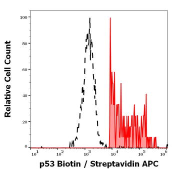
Flow cytometry intracellular staining pattern of human peripheral whole blood stained using anti-p53 (BP53-12) Biotin antibody (concentration in sample 1 μg/ml, Streptavidin APC).
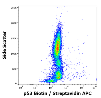
Flow cytometry intracellular staining pattern of human peripheral whole blood stained using anti-p53 (BP53-12) Biotin antibody (concentration in sample 1 μg/ml, Streptavidin APC).
Documents Download
Datasheet
Product Information
Request a Document
Protocol Information
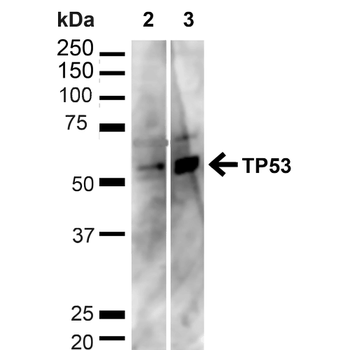
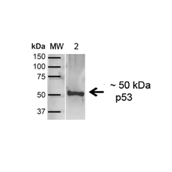
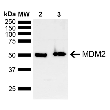
WB
Western Blot (IB, immunoblot)
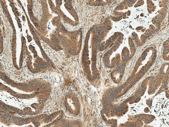
IHC-P
Immunohistochemistry Paraffin
FC
Flow Cytometry
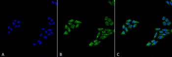
ICC
Immunocytochemistry
ELISA
Enzyme-linked Immunosorbent Assay (EIA)
IP
Immunoprecipitation
p53 Antibody (Biotin) (orb2653067)

- 0.0
Based on 0 reviews
Participating in our Biorbyt product reviews program enables you to support fellow scientists by sharing your firsthand experience with our products.
Login to Submit a ReviewAvailable Sizes
Select a size below
Free Secondary Antibody (20 ul)0/0
Please add an antibody product to your cart first.