You have no items in your shopping cart.
Description
Research Area
Immunology & Inflammation
Images & Validation
−Item 1 of 4
| Tested Applications | FC |
|---|---|
| Reactivity | Human |
| Application Notes |
Key Properties
−| Antibody Type | Primary Antibody |
|---|---|
| Clonality | Monoclonal |
| Isotype | Mouse IgG1 |
| Clone No. | HIT2 |
| Immunogen | Human thymocytes in foetus |
| Target | CD38 |
| Purification | Purified antibody is conjugated with fluorescein isothiocyanate (FITC) under optimum conditions and unconjugated antibody and free fluorochrome are removed by size-exclusion chromatography. |
| Conjugation | FITC |
Storage & Handling
−| Storage | Store at 2-8°C. Protect from prolonged exposure to light. Do not freeze. |
|---|---|
| Buffer/Preservatives | Stabilizing phosphate buffered saline (PBS), pH 7.4, 15 mM sodium azide |
| Expiration Date | 12 months from date of receipt. |
| Disclaimer | For research use only |
Alternative Names
−ADPRC1, cADPr hydrolase 1, T10, NAD(+) nucleosidase, ADP-ribosyl cyclase 1
Similar Products
−- Item 1 of 4

- Item 1 of 1
CD38/ADPRC 1 Human Monoclonal Antibody (FITC) [orb2970314]
ELISA, FC
Human
Human
Monoclonal
FITC
100 T, 50 T - Item 1 of 1
- Item 1 of 1


Quality Guarantee
Explore bioreagents carefree to elevate your research. All our products are rigorously tested for performance. If a product does not perform as described on its datasheet, our scientific support team will provide expert troubleshooting, a prompt replacement, or a refund. For full details, please see our Terms & Conditions and Buying Guide. Contact us at [email protected].
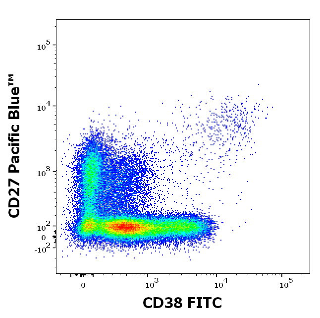
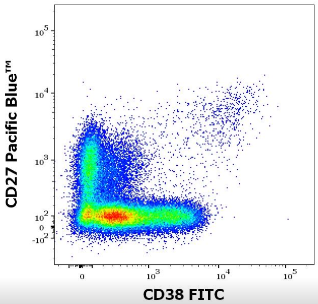
Staining pattern of Anti-human CD38 FITC antibody (clone HIT2) in dot-plot fluorescence visualization (B cell gate). Analysis of the antibody staining profile was performed on blood leukocytes isolated from buffy coats. Mouse monoclonal anti-human CD38 FITC antibody (clone HIT2) in concentration 0.5 µg/ml in stained stained blood sample (2 x 10^6 cells) and Mouse monoclonal anti-human CD27 Pacific Blue™ antibody (clone LT27) was used in concentration 2 µg/ml, respectively.
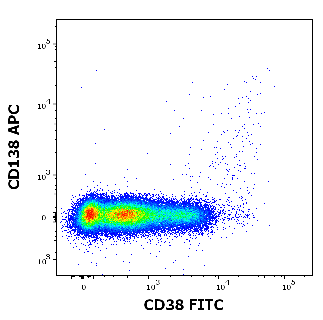
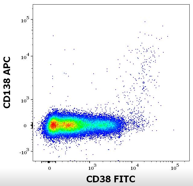
Staining pattern of Anti-human CD38 FITC antibody (clone HIT2) in dot-plot fluorescence visualization (B cell gate). Analysis of the antibody staining profile was performed on blood leukocytes isolated from buffy coats. Mouse monoclonal anti-human CD38 FITC antibody (clone HIT2) in concentration 0.5 µg/ml in stained stained blood sample (2 x 10^6 cells) and Mouse monoclonal anti-human CD138 APC antibody (clone MI15) in concentration 2 µg/ml, respectively.
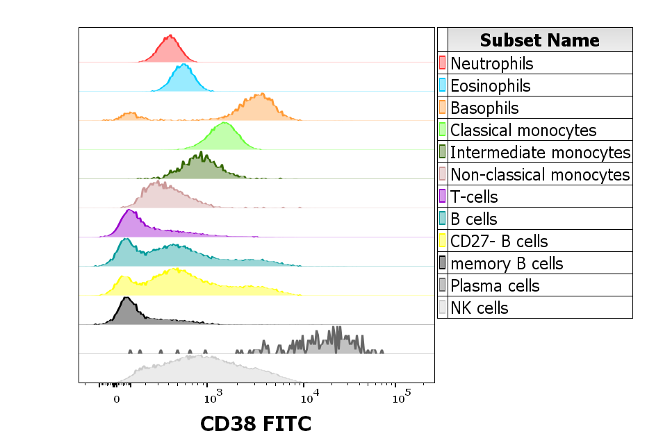
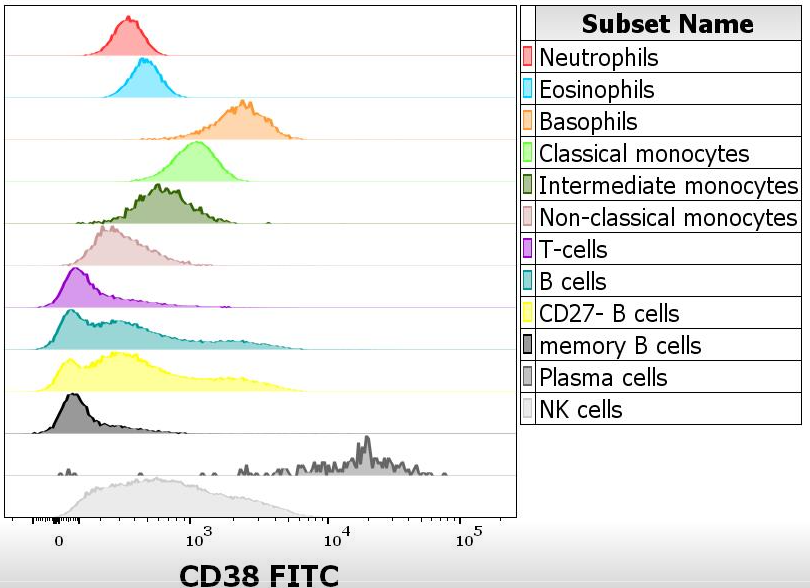
Reactivity of Anti-human CD38 FITC antibody (clone HIT2) on human peripheral leukocytes. Analysis of the antibody staining profile was performed on blood leukocytes isolated from buffy coats. Suspension of blood leukocytes (2 x 10^6 cells) was added to the mixture of CD38 FITC antibody (clone HIT2, 0.5 µg/ml in stained blood sample), backbone antibody conjugates and Monocyte Blocking Buffer, vortexed and incubated for 20 min. Stained sample was fixed with 2 ml of 10× diluted EXCELLYSE Easy solution for 10 min. Finally, samples were centrifuged (670 g, 5 min.), supernatant removed and the cell pellet was resuspended in 200 µl of PBS for acquisition.
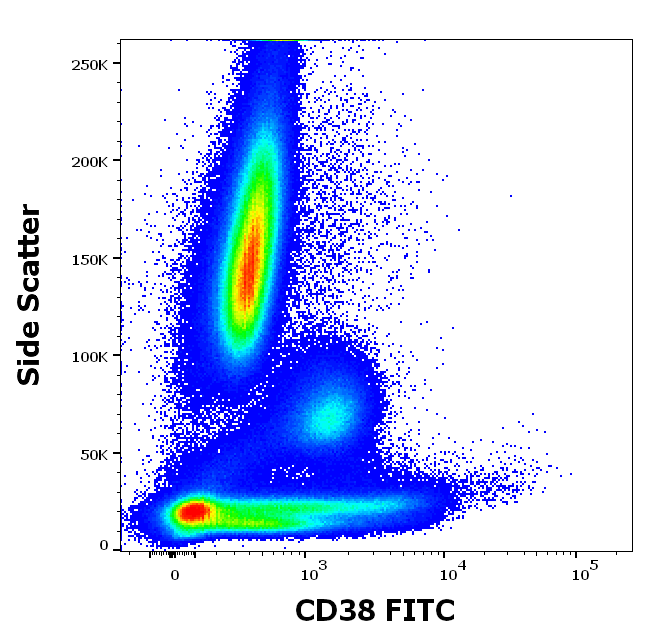
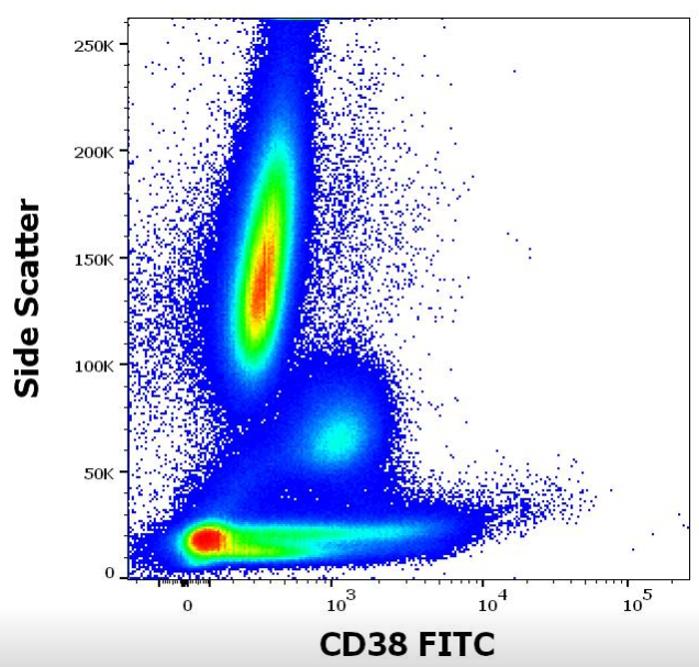
Anti-human CD38 FITC antibody (clone HIT2) works in flow cytometry application. Analysis of the antibody staining profile was performed on blood leukocytes isolated from buffy coats. Mouse monoclonal anti-human CD38 FITC antibody (clone HIT2) was used in concentration 0.5 µg/ml in stained blood sample (2 x 10^6 cells).
Documents Download
Datasheet
Product Information
Request a Document
CD38 Antibody (FITC) (orb43853)

- 0.0
Based on 0 reviews
Participating in our Biorbyt product reviews program enables you to support fellow scientists by sharing your firsthand experience with our products.
Login to Submit a ReviewAvailable Sizes
Select a size below
Free Secondary Antibody (20 ul)0/0
Please add an antibody product to your cart first.