You have no items in your shopping cart.
Description
Research Area
Immunology & Inflammation
Images & Validation
−Item 1 of 4
| Tested Applications | FC, IHC-Fr, IP |
|---|---|
| Reactivity | Human, Primate |
| Application Notes |
Key Properties
−| Antibody Type | Primary Antibody |
|---|---|
| Clonality | Monoclonal |
| Isotype | Mouse IgG1 kappa |
| Clone No. | 3G8 |
| Immunogen | Human neutrophils |
| Target | CD16 |
| Purification | Purified by protein-A affinity chromatography. |
| Conjugation | Unconjugated |
Storage & Handling
−| Storage | Maintain refrigerated at 2-8°C for up to 2 weeks. For long term storage store at -20°C in small aliquots to prevent freeze-thaw cycles. |
|---|---|
| Buffer/Preservatives | Phosphate buffered saline (PBS), pH 7.4, 15 mM sodium azide |
| Concentration | 1 mg/ml |
| Expiration Date | 12 months from date of receipt. |
| Disclaimer | For research use only |
Alternative Names
−FcgammaRIII, IGFR3, FCRIII
Similar Products
−- Item 1 of 7
- Item 1 of 4
CD16 Rabbit Polyclonal Antibody [orb101668]
WB
Human
Mouse, Rat
Rabbit
Polyclonal
Unconjugated
50 μl, 100 μl, 200 μl - Item 1 of 5
- Item 1 of 5
- Item 1 of 5
CD16/FCGR3A Rabbit Polyclonal Antibody [orb402246]
ELISA, FC, ICC, IF, IHC
Human
Rabbit
Polyclonal
Unconjugated
100 μg


Quality Guarantee
Explore bioreagents carefree to elevate your research. All our products are rigorously tested for performance. If a product does not perform as described on its datasheet, our scientific support team will provide expert troubleshooting, a prompt replacement, or a refund. For full details, please see our Terms & Conditions and Buying Guide. Contact us at [email protected].

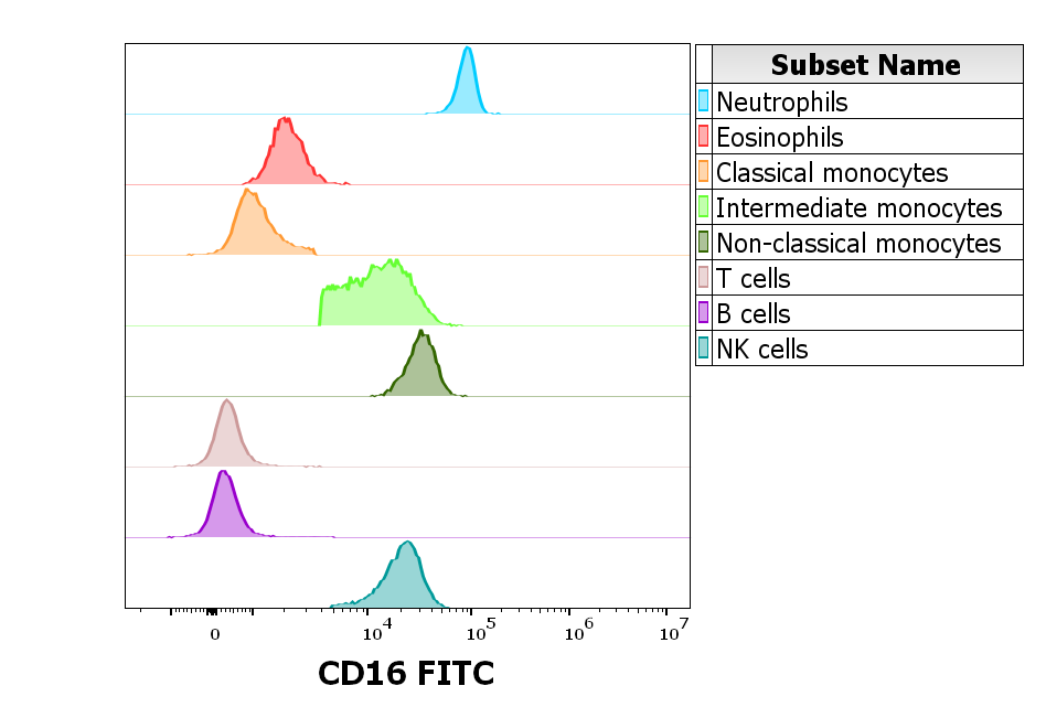
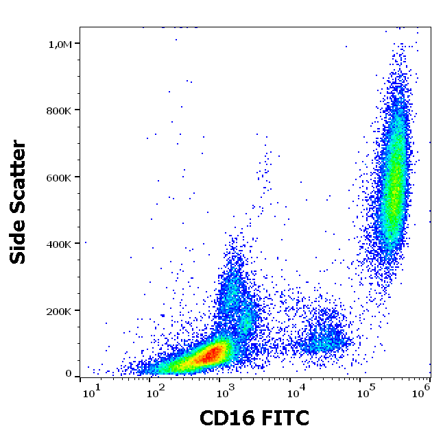
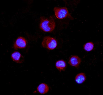
Expression profiling on peripheral blood subsets using anti-human CD16 purified antibody (clone 3G8). HCDM CDMaps standardized procedures were used for cell isolation and surface staining of blood leukocytes, with the modification of staining protocol using cytometry test tubes. Suspension of blood leukocytes isolated from buffy coats (2 x 10^6 cells) with residual erythrocytes lysed with 10× diluted EXCELLYSE Live solution was added to the mixture of anti-human CD16 purified antibody (clone 3G8, 0.5 µg/ml in stained blood sample) and Monocyte Blocking Buffer, vortexed and incubated for 20 min. Next, samples were centrifuged (670 g, 5 min.), supernatant removed and secondary antibody (GAM PE) was added to sample, vortexed and incubated for 20 min. Next, samples were washed twice (2 ml PBS, 670 g, 5 min.) and then optimized backbone antibody panels (HLDA Innate and HLDA Adaptive) were added to test tubes, vortexed and incubated for 20 min. Next, samples are fixed with 2 ml of 10× diluted EXCELLYSE Easy solution and incubated for 10 min. Finally, samples were centrifuged (670 g, 5 min.), supernatant removed and the cell pellet was resuspended in 200 µl of PBS for acquisition.
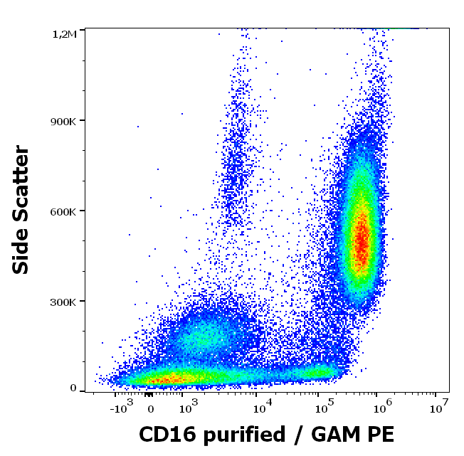
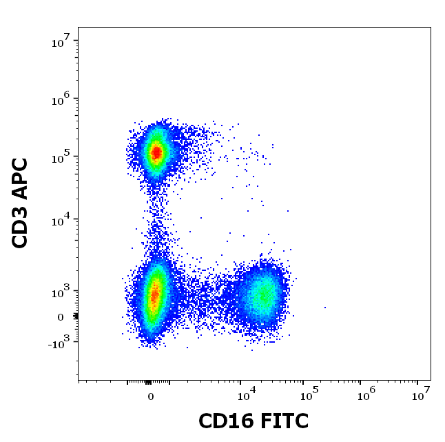
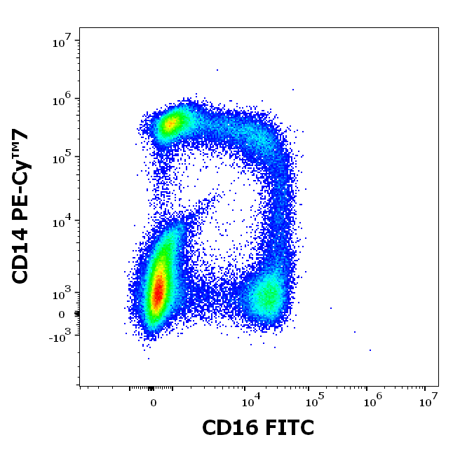
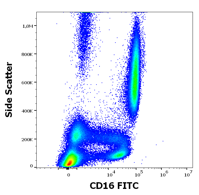
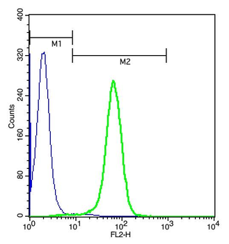
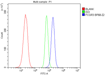
Anti-human CD16 purified antibody (clone 3G8) works in flow cytometry application. Analysis of the antibody staining profile was performed on blood leukocytes isolated from buffy coats. HCDM CDMaps standardized procedures were used for cell isolation and surface staining of blood leukocytes, with the modification of staining protocol using cytometry test tubes. Mouse monoclonal anti-human CD16 purified antibody (clone 3G8) was used in concentration 0.5 µg/ml in stained blood sample (2 x 10^6 cells).
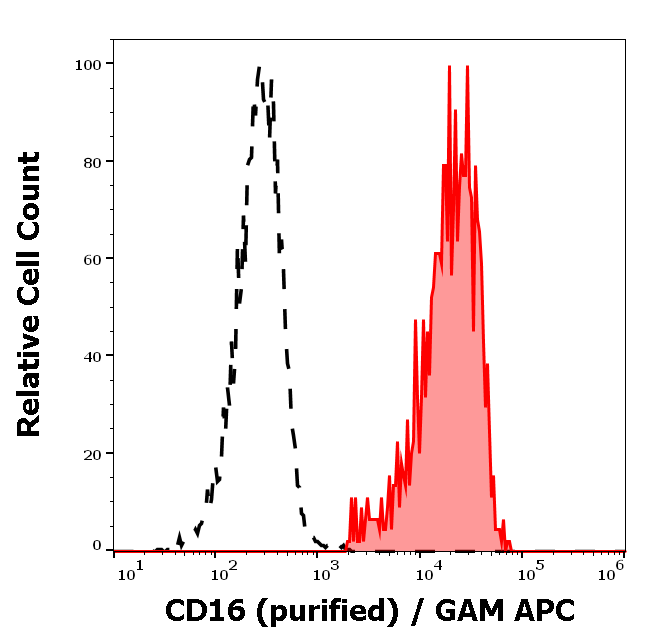
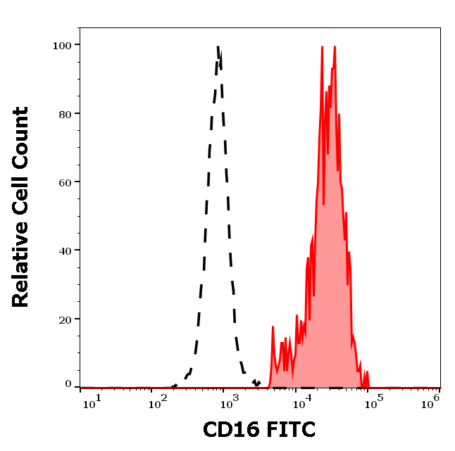
Separation of human CD16 positive lymphocytes (red-filled) from CD16 negative lymphocytes (black-dashed) in flow cytometry analysis (surface staining) of peripheral whole blood stained using anti-human CD16 (3G8) purified antibody (concentration in sample 2 µg/ml, GAM APC).
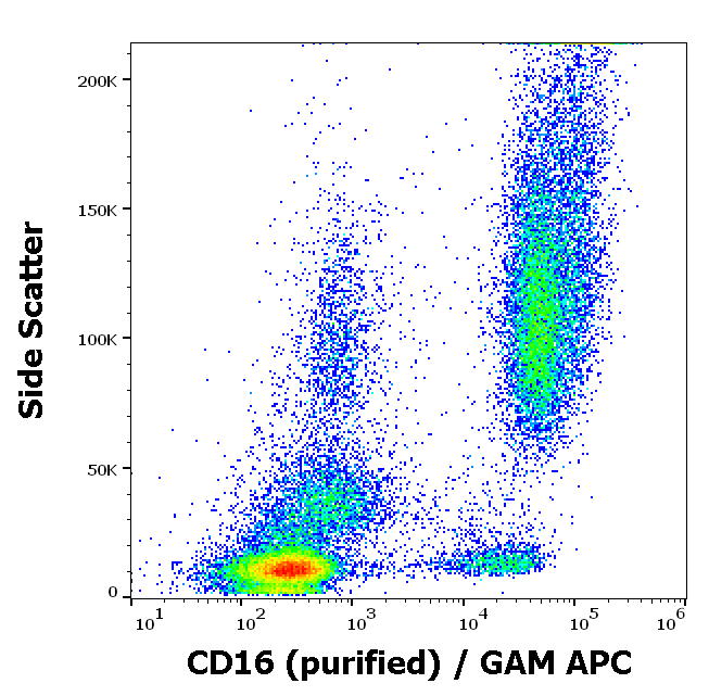
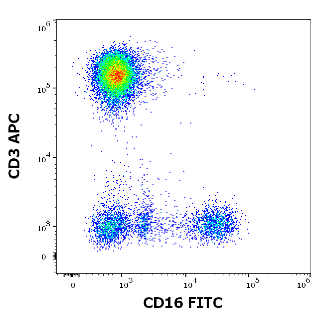
Flow cytometry surface staining pattern of human peripheral whole blood stained using anti-human CD16 (3G8) purified antibody (concentration in sample 2 µg/ml, GAM APC).
Documents Download
Datasheet
Product Information
Request a Document
Protocol Information
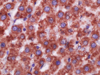
IHC-Fr
Immunohistochemistry Frozen
FC
Flow Cytometry
IP
Immunoprecipitation
CD16 Antibody (orb44653)

- 0.0
Based on 0 reviews
Participating in our Biorbyt product reviews program enables you to support fellow scientists by sharing your firsthand experience with our products.
Login to Submit a ReviewAvailable Sizes
Select a size below
Free Secondary Antibody (20 ul)0/0
Please add an antibody product to your cart first.