You have no items in your shopping cart.
Description
Research Area
Epigenetics & Chromatin
Images & Validation
−Item 1 of 3
| Tested Applications | FC, IF, IHC, WB |
|---|---|
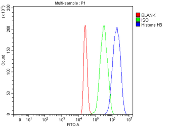
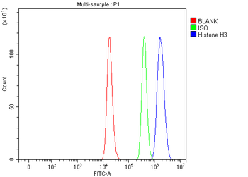
| Dilution Range | WB: 1:500-1000, IHC-P: 1:100-200, IF/ICC: 1:100-500, FACS: 1:100-200 |
| Reactivity | Bovine, Gallus, Human, Mouse, Porcine, Rat, Zebrafish |
Key Properties
−| Antibody Type | Primary Antibody |
|---|---|
| Host | Rabbit |
| Clonality | Polyclonal |
| Immunogen | KLH-conjugated synthetic peptide encompassing a sequence within the N-term region of human Histone H3. The exact sequence is proprietary. |
| Target | HIST1H3A |
| Purification | The antibody was purified by immunogen affinity chromatography. |
| Conjugation | Unconjugated |
Storage & Handling
−| Storage | Maintain refrigerated at 2-8°C for up to 2 weeks. For long term storage store at -20°C in small aliquots to prevent freeze-thaw cycles. |
|---|---|
| Form/Appearance | Liquid |
| Buffer/Preservatives | 0.42% Potassium phosphate, 0.87% Sodium chloride, pH 7.3, 30% glycerol, and 0.01% sodium azide. |
| Expiration Date | 12 months from date of receipt. |
| Disclaimer | For research use only |
Alternative Names
−HIST1H3A; H3FA; HIST1H3B; H3FL; HIST1H3C; H3FC; HIST1H3D; H3FB; HIST1H3E; H3FD; HIST1H3F; H3FI; HIST1H3G; H3FH; HIST1H3H; H3FK; HIST1H3I; H3FF; HIST1H3J; H3FJ; Histone H3.1; Histone H3/a; Histone H3/b; Histone H3/c; Histone H3/d; Histone H3/f; Histone H3/h; Histone H3/i; Histone H3/j; Histone H3/k; Histone H3/l; HIST2H3A; HIST2H3C; H3F2; H3FM; HIST2H3D; Histone H3.2; Histone H3/m; Histone H3/o; H3F3A; H3.3A; H3F3; PP781; H3F3B; H3.3B; Histone H3.3
Similar Products
−- Item 1 of 48
Histone H3 Rabbit Polyclonal Antibody (Nuclear Loading Control) [orb10805]
FC, ICC, IF, IHC-Fr, IHC-P, WB
Hamster, Insect, Yeast
Human, Mouse, Rat
Rabbit
Polyclonal
Unconjugated
100 μl, 500 μl, 200 μl, 1 ml - Item 1 of 25
Histone H3 HIST1H3A/B/C/D/E/F/G/H/I/J Rabbit Polyclonal Antibody [orb614120]
ELISA, FC, ICC, IF, IHC, WB
Human, Mouse, Rat
Rabbit
Polyclonal
Unconjugated
100 μg - Item 1 of 14
Histone H3.1 Rabbit Polyclonal Antibody [orb184149]
IF, IHC-Fr, IHC-P, WB
Bovine, Equine, Porcine, Primate, Rabbit, Sheep
Human, Mouse, Rat
Rabbit
Polyclonal
Unconjugated
50 μl, 100 μl, 200 μl - Item 1 of 17
Histone H3 (Tri Methyl K4) Rabbit Polyclonal Antibody [orb1845]
ELISA, IF, IHC-Fr, IHC-P, WB
Mouse, Rat
Human, Mouse, Rat
Rabbit
Polyclonal
Unconjugated
50 μl, 100 μl - Item 1 of 13
Histone H3.3B Rabbit Polyclonal Antibody [orb556958]
ICC, IHC-P, WB
Human, Mouse, Rat
Rabbit
Polyclonal
Unconjugated
100 μl


Quality Guarantee
Explore bioreagents carefree to elevate your research. All our products are rigorously tested for performance. If a product does not perform as described on its datasheet, our scientific support team will provide expert troubleshooting, a prompt replacement, or a refund. For full details, please see our Terms & Conditions and Buying Guide. Contact us at [email protected].
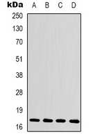
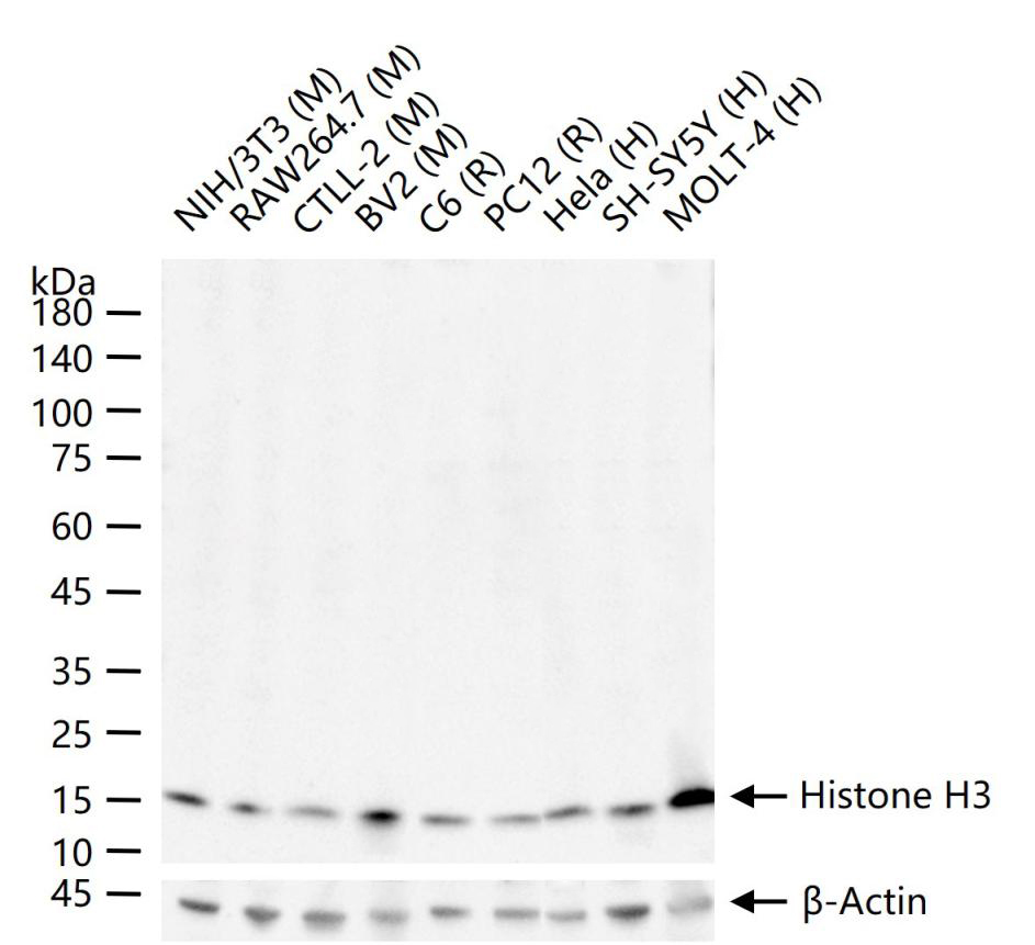
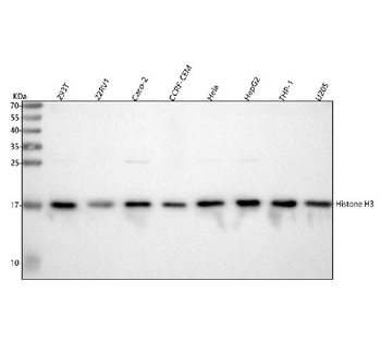
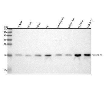
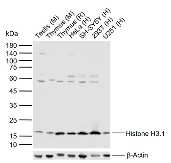
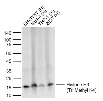
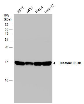
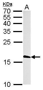
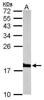
Western blot analysis of Histone H3 expression in MCF7 (A), Raw264.7 (B), mouse heart (C), rat heart (D) whole cell lysates. (Predicted band size: 15 kD; Observed band size: 17 kD)
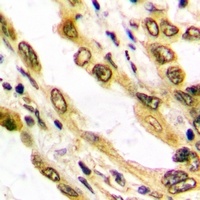
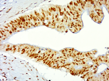
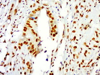
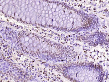
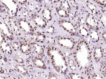
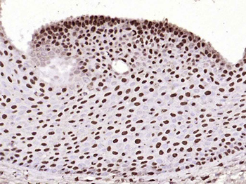
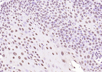
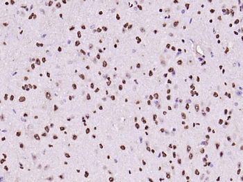
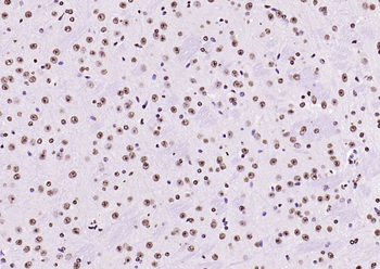
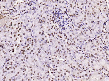
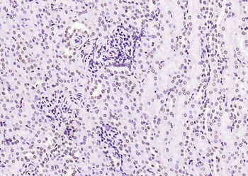
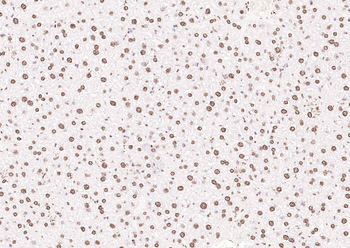
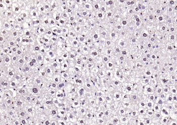
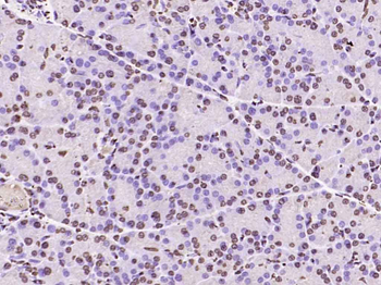
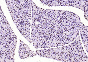
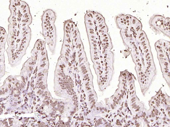
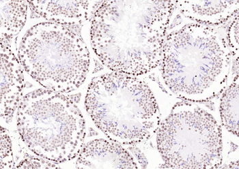
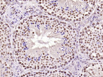
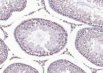
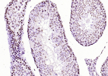
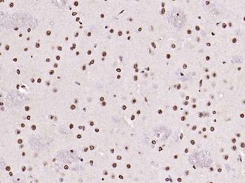
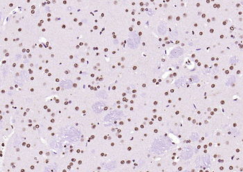
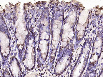
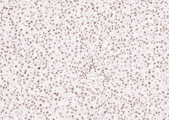
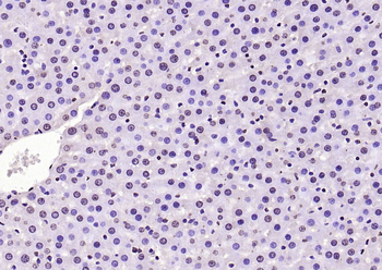
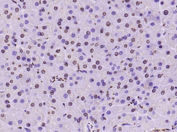
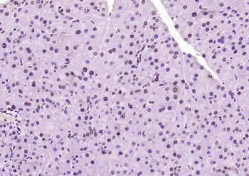
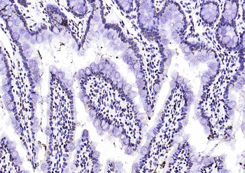
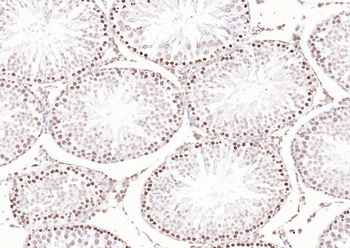
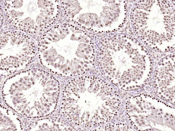
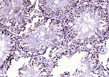
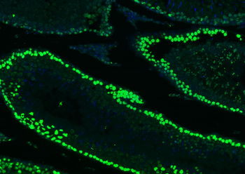
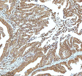
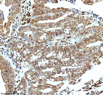
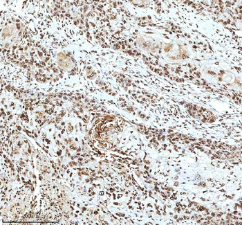
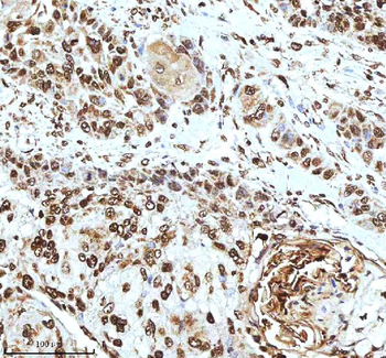
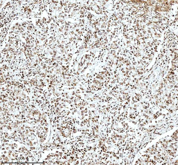
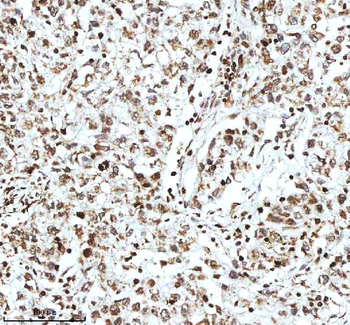
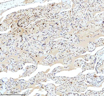
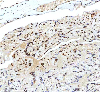
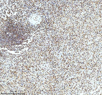
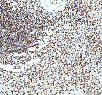
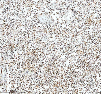
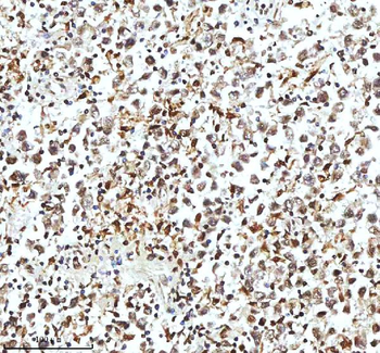
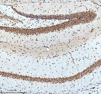
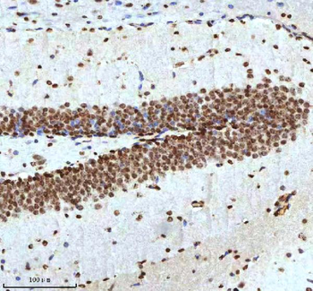
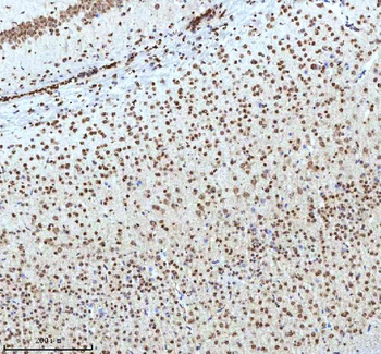
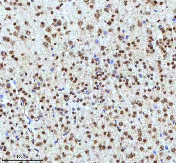
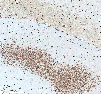
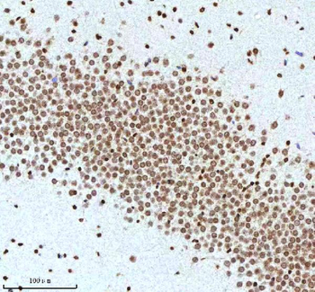
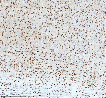
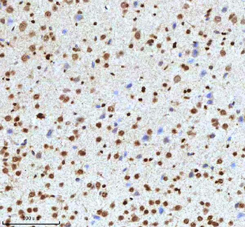
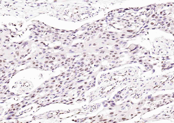
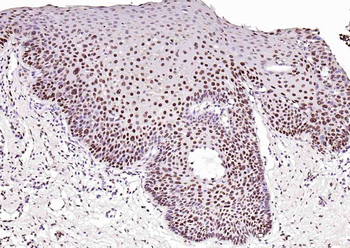
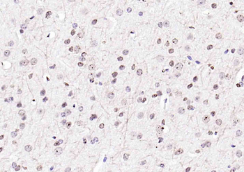
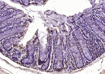
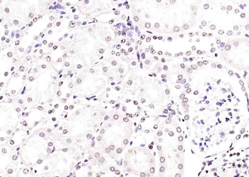
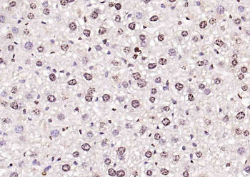
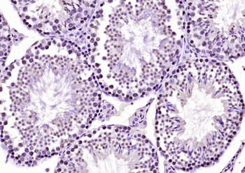
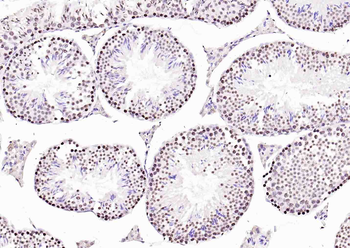
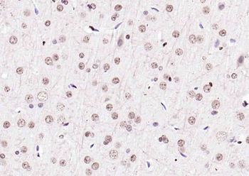
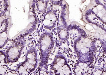
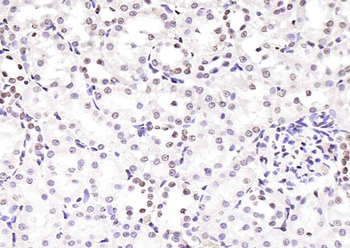
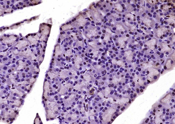
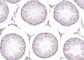
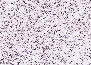
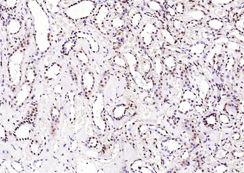
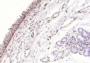
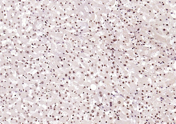
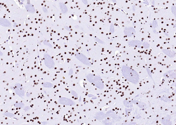
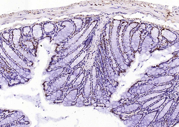
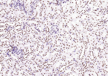
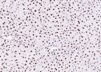
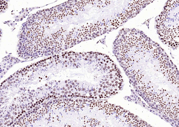
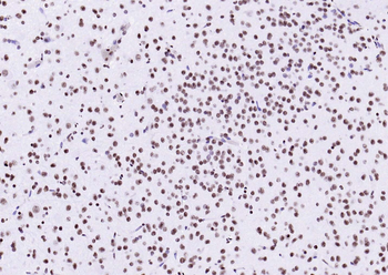
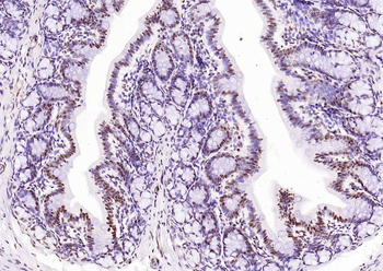
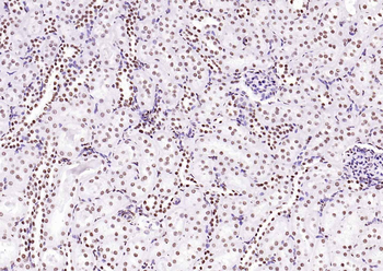
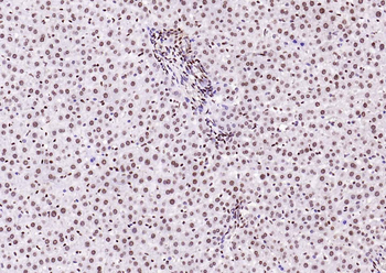
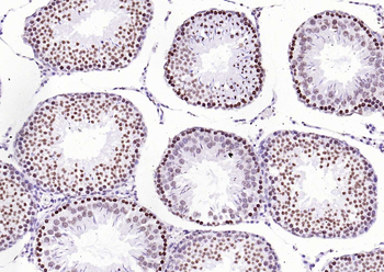
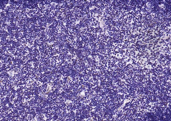
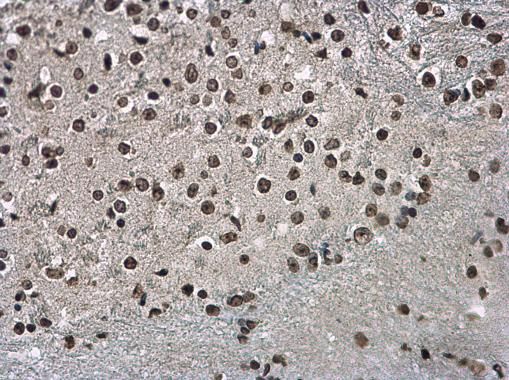
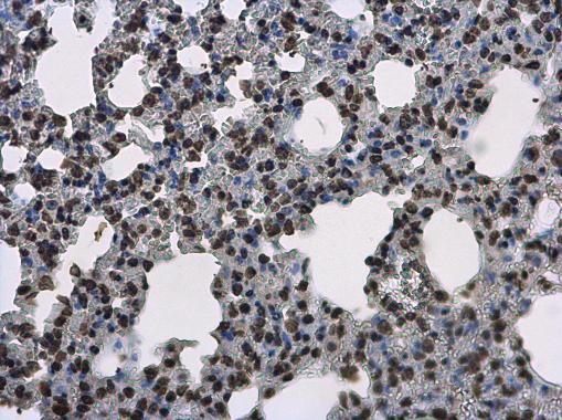
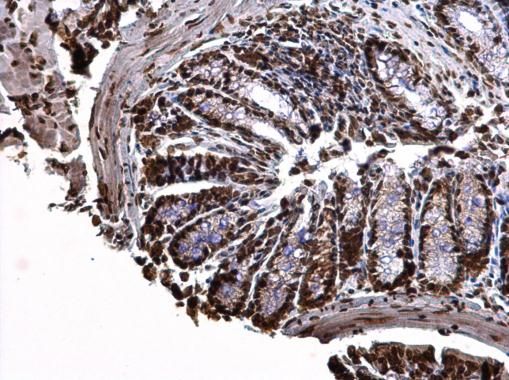
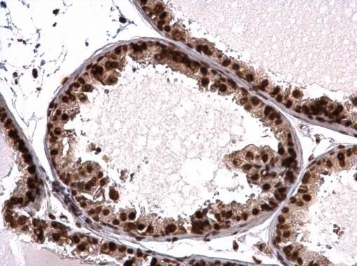
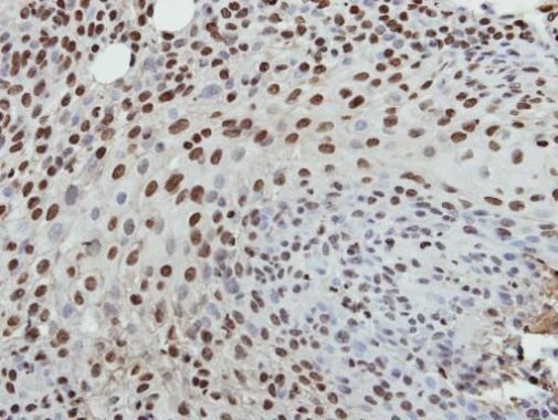
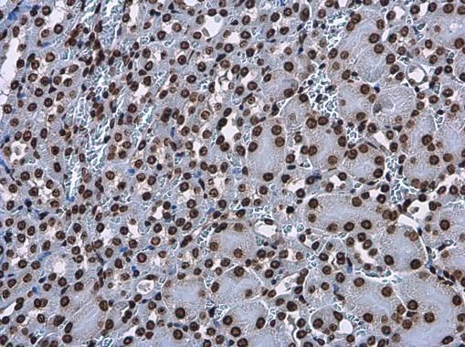
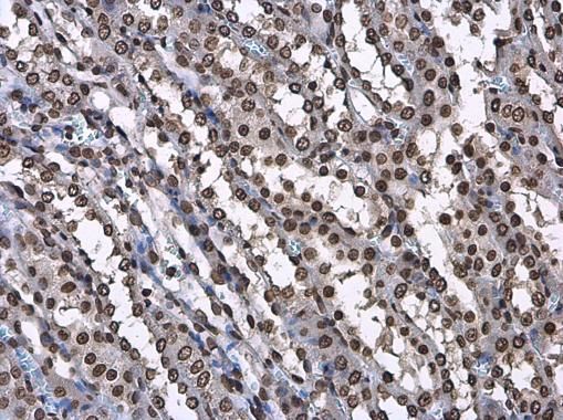
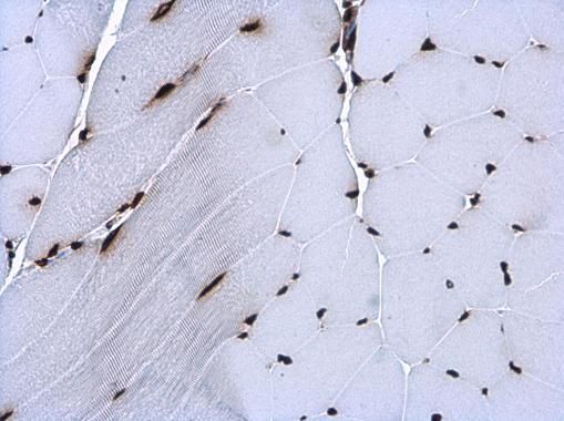
Immunohistochemical analysis of Histone H3 staining in human lung cancer formalin fixed paraffin embedded tissue section. The section was pre-treated using heat mediated antigen retrieval with sodium citrate buffer (pH 6.0). The section was then incubated with the antibody at room temperature and detected using an HRP conjugated compact polymer system. DAB was used as the chromogen. The section was then counterstained with haematoxylin and mounted with DPX.
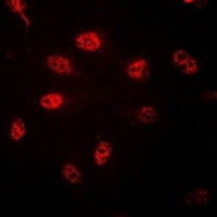
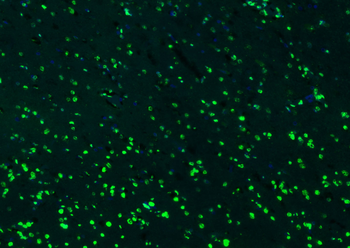
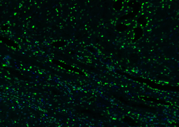
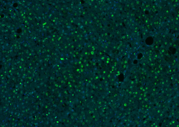
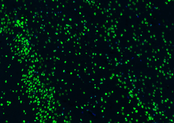
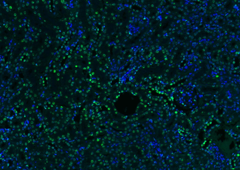
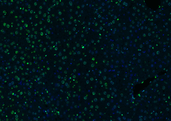
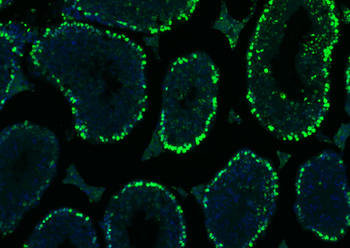
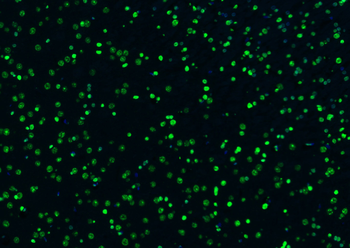
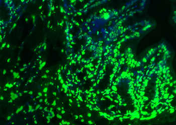
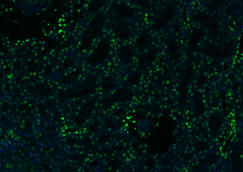
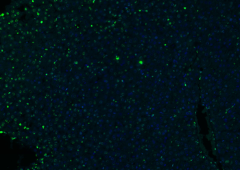
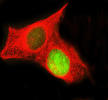
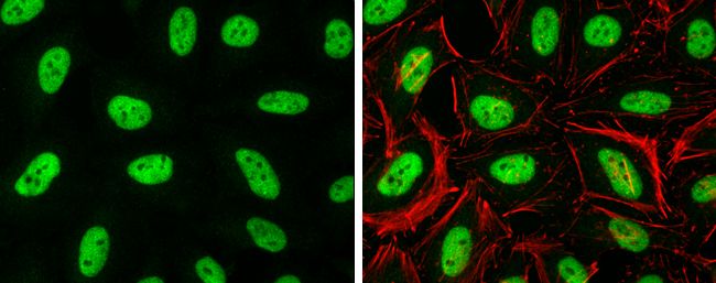
Immunofluorescent analysis of Histone H3 staining in HeLa cells. Formalin-fixed cells were permeabilized with 0.1% Triton X-100 in TBS for 5-10 minutes and blocked with 3% BSA-PBS for 30 minutes at room temperature. Cells were probed with the primary antibody in 3% BSA-PBS and incubated overnight at 4 °C in a hidified chamber. Cells were washed with PBST and incubated with a DyLight 594-conjugated secondary antibody (red) in PBS at room temperature in the dark.
Quick Database Links
UniProt Details
− No UniProt data available
NCBI Gene Details
− No NCBI Gene data available
Documents Download
Datasheet
Product Information
Request a Document
Protocol Information
WB
Western Blot (IB, immunoblot)
IHC
Immunohistochemistry
FC
Flow Cytometry
IF
Immunofluorescence
Histone H3 Rabbit Polyclonal Antibody (orb338856)

- 0.0
Based on 0 reviews
Participating in our Biorbyt product reviews program enables you to support fellow scientists by sharing your firsthand experience with our products.
Login to Submit a ReviewAvailable Sizes
Select a size below
Choose Conjugation or Carrier Free Version
Free Secondary Antibody (20 ul)0/0
Please add an antibody product to your cart first.