You have no items in your shopping cart.
Description
Images & Validation
−Item 1 of 2
| Tested Applications | ICC, IP, WB |
|---|---|
| Reactivity | All |
| Application Notes |
Key Properties
−| Antibody Type | Primary Antibody |
|---|---|
| Clonality | Polyclonal |
| Isotype | Rabbit polyclonal |
| Immunogen | EGFP, a native full-length protein |
| Target | GFP |
| Purification | Purified by protein-A affinity chromatography. |
| Conjugation | Unconjugated |
Storage & Handling
−| Storage | Maintain refrigerated at 2-8°C for up to 2 weeks. For long term storage store at -20°C in small aliquots to prevent freeze-thaw cycles. |
|---|---|
| Buffer/Preservatives | Phosphate buffered saline (PBS), pH 7.4, 15 mM sodium azide |
| Concentration | 1 mg/ml |
| Expiration Date | 12 months from date of receipt. |
| Disclaimer | For research use only |
Alternative Names
−Green fluorescent protein, EGFP, EYFP
Similar Products
−- Item 1 of 28
- Item 1 of 28
- Item 1 of 28
- Item 1 of 7
- Item 1 of 6


Quality Guarantee
Explore bioreagents carefree to elevate your research. All our products are rigorously tested for performance. If a product does not perform as described on its datasheet, our scientific support team will provide expert troubleshooting, a prompt replacement, or a refund. For full details, please see our Terms & Conditions and Buying Guide. Contact us at [email protected].
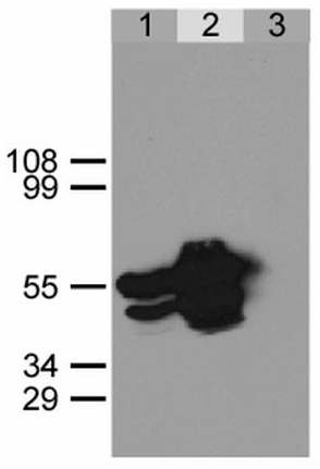
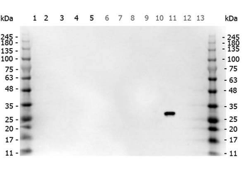
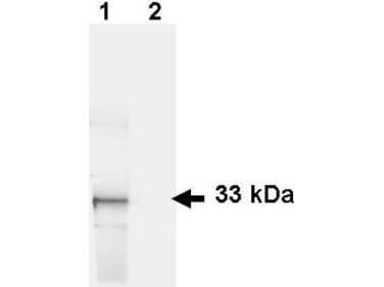
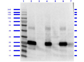
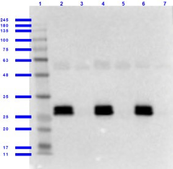
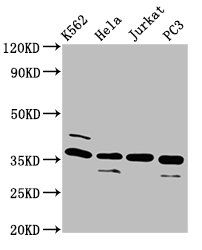
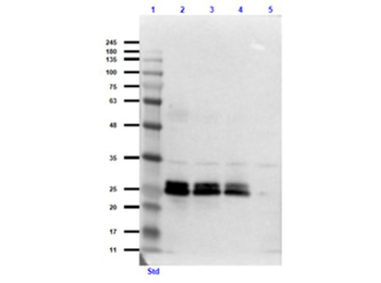
Immunoprecipitation of GFP-NLS from HEK293 cells using anti-GFP antibody. HEK293 cells were transfected with expression construct encoding GFP-NLS protein. Twenty hours post transfection cells were lysed in non-denaturating conditions (Lysis buffer: 20 mM Tris, pH 7.5, 100 mM NaCl, 0.5% Triton X-100, inhibitors of proteases). Aliquots of cell lysate were immunoprecipitated using a polyclonal anti-GFP antibody (lane 2) or a pre-immune rabbit serum (lane 3). Immunoprecipitates together with a sample of the cell lysate (lane 1) were separated on SDS-PAGE polyacrylamide gel and immunoblotted with the anti-GFP antibody. The positions of molecular weight markers in kDa are indicated at the left.
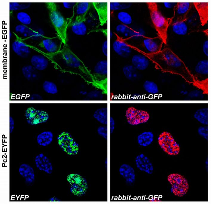
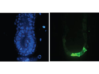
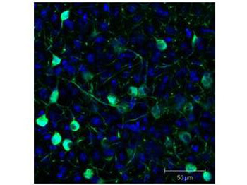
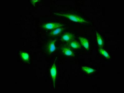
Immunocytochemistry staining (confocal microscopy) of COS-7 cells transfected with expression constructs encoding membrane-tethered EGFP (membrane-EGFP; top) or nuclear Polycomb 2-EYFP fusion protein (Pc2-EYFP; bottom). The natural fluorescence of the produced proteins is shown in the green channel (left), polyclonal anti-GFP antibody signal was detected in the red channel (right). The system was carefully tested for overlap of these two optical channels and images were scanned separately in sequential scanning mode. The blue nuclear stain is also shown.
Documents Download
Datasheet
Product Information
Request a Document
Protocol Information
WB
Western Blot (IB, immunoblot)
ICC
Immunocytochemistry
IP
Immunoprecipitation
GFP Antibody (orb44764)

- 0.0
Based on 0 reviews
Participating in our Biorbyt product reviews program enables you to support fellow scientists by sharing your firsthand experience with our products.
Login to Submit a ReviewAvailable Sizes
Select a size below
Free Secondary Antibody (20 ul)0/0
Please add an antibody product to your cart first.