You have no items in your shopping cart.
Description
Images & Validation
−Item 1 of 6
| Tested Applications | ELISA, IF, IHC, WB |
|---|---|
| Dilution Range | ELISA: 1:20,000 - 1:120,000, IHC: 1:200 - 1:3,000, IF: 1:500 - 1:5,000, WB: 1:500 - 1:5,000 |
| Reactivity | Other |
| Application Notes |
Key Properties
−| Antibody Type | Primary Antibody |
|---|---|
| Host | Rabbit |
| Clonality | Polyclonal |
| Isotype | IgG |
| Immunogen | The immunogen is a Green Fluorescent Protein (GFP) fusion protein corresponding to the full length amino acid sequence (246aa) derived from the jellyfish Aequorea victoria. |
| Purity | Anti-GFP antibody was prepared from monospecific antiserum by immunoaffinity chromatography using Green Fluorescent Protein (Aequorea victoria) coupled to agarose beads followed by solid phase adsorption(s) to remove any unwanted reactivities. Assay by immunoelectrophoresis resulted in a single precipitin arc against anti-Rabbit Serum and purified and partially purified Green Fluorescent Protein (Aequorea victoria). No reaction was observed against Human, Mouse or Rat serum proteins. |
| Conjugation | Unconjugated |
Storage & Handling
−| Storage | Store Anti-GFP Antibody at -20° C prior to opening. Aliquot contents and freeze at -20° C or below for extended storage. Avoid cycles of freezing and thawing. Centrifuge product if not completely clear after standing at room temperature. GFP antibody is stable for several weeks at 4° C as an undiluted liquid. Dilute only prior to immediate use. |
|---|---|
| Form/Appearance | Liquid (sterile filtered) |
| Buffer/Preservatives | Preservative: 0.01% (w/v) Sodium Azide. Stabilizer: None; Buffer: 0.02 M Potassium Phosphate, 0.15 M Sodium Chloride, pH 7.2 |
| Concentration | 1.25 mg/mL |
| Expiration Date | 12 months from date of receipt. |
| Dry Ice Shipping | Please note: This product requires shipment on dry ice. A dry ice surcharge will apply. |
| Disclaimer | For research use only |
Alternative Names
−rabbit anti-GFP antibody, Green Fluorescent Protein, GFP antibody, Green Fluorescent Protein antibody, EGFP, enhanced Green Fluorescent Protein, Aequorea victoria, Jellyfish
Similar Products
−- Item 1 of 28
- Item 1 of 28
- Item 1 of 28
- Item 1 of 7
- Item 1 of 6


Quality Guarantee
Explore bioreagents carefree to elevate your research. All our products are rigorously tested for performance. If a product does not perform as described on its datasheet, our scientific support team will provide expert troubleshooting, a prompt replacement, or a refund. For full details, please see our Terms & Conditions and Buying Guide. Contact us at [email protected].
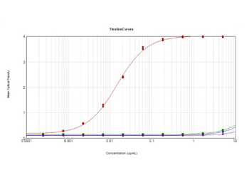
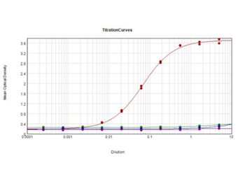
ELISA results of purified Rabbit Anti-GFP Antibody. Each well was coated in 10 µg of antigen GFP [Red Line], human IgG [Green Line], Mouse IgG [Blue Line], and Rat IgG [Purple Line]. The starting dilution of antibody was 5 µg/ml and the X-axis represents the Log10 of a 3-fold dilution. The titer is 1:67700. This titration is a 4-parameter curve fit where the IC50 is defined as the titer of the antibody. Assay performed using 1% Fish Gel, TMB Substrate, and Goat Anti-Rabbit IgG Antibody HRP (p/n orb347654).
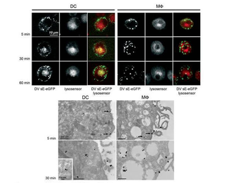
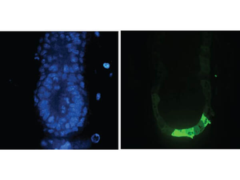
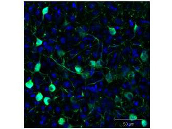
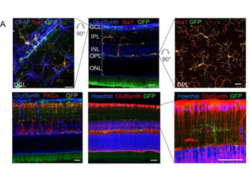
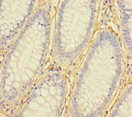
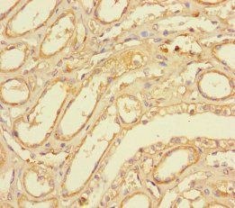
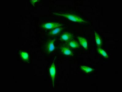
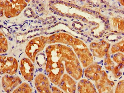
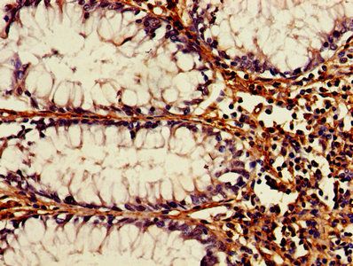
Immuno-microscopy of Rabbit anti-GFP antibody. Monocyte derived dendritic cells and dermal macrophages were challenged and directly visualized with eGFP labeled Dengue virus to localize sequestration of virus particles in the different cells (upper). The location of the GFP was confirmed by TEM (lower magnified view) using Biorbyt rabbit anti GFP Primary antibody (1:200) and a gold labeled secondary antibody.
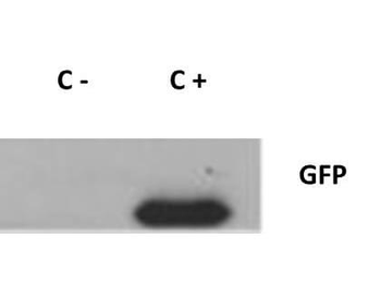
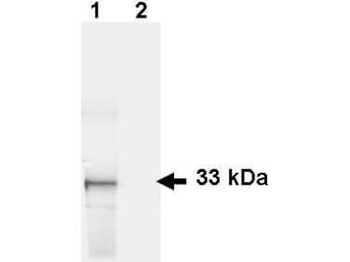
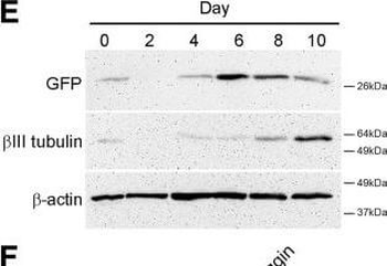
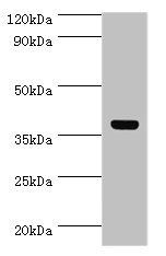
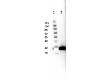
Western Blot of Rabbit anti-GFP antibody. Lane 1: 293FT cells transfected with CDK4 dominant negative (C-). Lane 2: 293FT cells poitive control (C +). Load: 25 µg per lane. Primary antibody: GFP antibody at 1:400 for overnight at 4°C. Secondary antibody: IRDye800™ rabbit secondary antibody at 1:10000 for 45 min at RT. Block: 5% BLOTTO overnight at 4°C. Predicted/Observed size: 27 kDa for GFP.
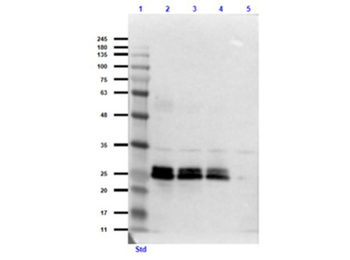
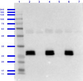
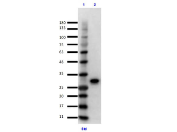
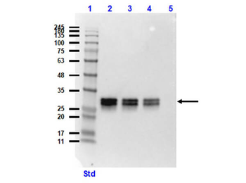
Western Blot of Rabbit Anti-GFP Antibody. Lane 1: Opal Prestained Molecular Weight Ladder. Lane 2: GFP (p/n orb345957) / HeLa Lysate (p/n orb348668) [0.1 µg/10.0 µg]. Lane 3: GFP (p/n orb345957) / HeLa Lysate (p/n orb348668) [0.05 µg/10.0 µg]. Lane 4: GFP (p/n orb345957) / HeLa Lysate (p/n orb348668) [0.03 µg/10.0 µg]. Lane 5: HeLa Lysate (p/n orb348668) [10.0 µg]. Primary Antibody: Rabbit Anti-GFP Antibody at 1.0 µg/ml overnight at 2-8°C. Secondary Antibody: Goat Anti-Rabbit IgG MX 10 Peroxidase (p/n orb347654) at 1:70000 for 30 mins at RT. Block: Blocking Buffer for Fluorescent Western Blotting (p/n orb348637) for 60 mins at RT. Expect: ~27kDa.
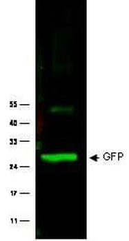
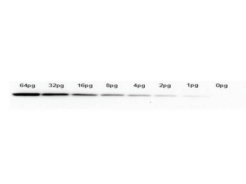
Western Blot of Rabbit anti-GFP antibody. Lane 1: Wild type GFP (0.1 µg) was used to spike HeLa whole cell lysate. Lane 2: none. Load: 30 µg per lane. Primary antibody: GFP antibody at 1:1000 for overnight at 4°C. Secondary antibody: IRDye800™ Goat-a-Rabbit IgG [H&L] MX10 at 1:10000 for 45 min at RT. Block: 5% BLOTTO in PBS overnight at 4°C. Predicted/Observed size: 27 kDa for epitope tag GFP. Other band(s): none.
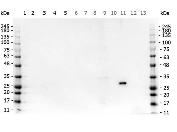
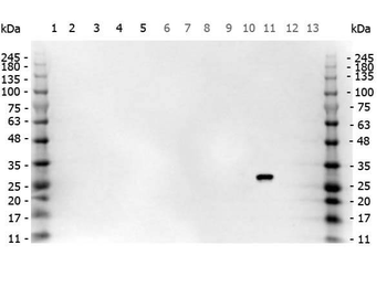
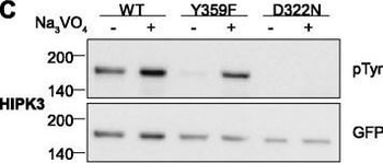
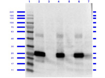
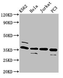
Western Blot of Rabbit anti-GFP antibody. Marker: Opal Pre-stained ladder. Lane 1: HEK293 lysate (p/n orb348669). Lane 2: HeLa Lysate (p/n orb348668). Lane 3: CHO/K1 Lysate. Lane 4: MDA-MB-231 (p/n orb348700). Lane 5: A431 Lysate (p/n orb348665). Lane 6: Jurkat Lysate (p/n orb348674). Lane 7: NIH/3T3 Lysate (p/n orb348714). Lane 8: E-coli HCP Control (p/n orb342262). Lane 9: FLAG Positive Control Lysate (p/n orb348661). Lane 10: Red Fluorescent Protein (p/n orb345960). Lane 11: Green Fluorescent Protein (p/n orb345957). Lane 12: Glutathione-S-Transferase Protein (p/n orb345956). Lane 13: Maltose Binding Protein. Load: 10 µg of lysate or 50 ng of purified protein per lane. Primary antibody: GFP antibody at 1 ug/ml overnight at 4C. Secondary antibody: Peroxidase rabbit secondary antibody (p/n orb347654) at 1:30000 for 60 min at RT. Blocking Buffer: 1% Casein-TTBS for 30 min at RT. Predicted/Observed size: 30 kDa for GFP.
Quick Database Links
UniProt
UniProt Details
− No UniProt data available
Documents Download
Datasheet
Product Information
Request a Document
Protocol Information
WB
Western Blot (IB, immunoblot)
IHC
Immunohistochemistry
IF
Immunofluorescence
ELISA
Enzyme-linked Immunosorbent Assay (EIA)
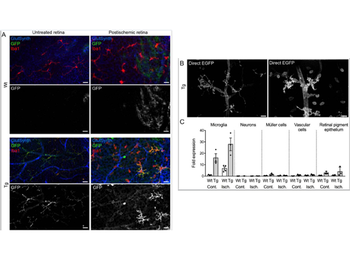
Widowati, Esti Wahyu et al. Functional characterization of DYRK1A missense variants associated with a syndromic form of intellectual deficiency and autism Biol Open, 7, (2018)
GFP Antibody (orb345367)

- 0.0
Based on 0 reviews
Participating in our Biorbyt product reviews program enables you to support fellow scientists by sharing your firsthand experience with our products.
Login to Submit a ReviewAvailable Sizes
Select a size below
Choose Conjugation or Carrier Free Version
Free Secondary Antibody (20 ul)0/0
Please add an antibody product to your cart first.