You have no items in your shopping cart.
Description
Research Area
Signal Transduction
Images & Validation
−Item 1 of 4
| Tested Applications | ICC, IHC-P, IP, WB |
|---|---|
| Reactivity | Human, Mouse |
| Application Notes |
Key Properties
−| Antibody Type | Primary Antibody |
|---|---|
| Clonality | Monoclonal |
| Isotype | Mouse IgG2b |
| Clone No. | FYN-01 |
| Immunogen | Bacterially expressed recombinant fragment of human Fyn (aa 7-176). |
| Target | FYN |
| Purification | Purified by protein-A affinity chromatography. |
| Conjugation | Unconjugated |
Storage & Handling
−| Storage | Maintain refrigerated at 2-8°C for up to 2 weeks. For long term storage store at -20°C in small aliquots to prevent freeze-thaw cycles. |
|---|---|
| Buffer/Preservatives | Phosphate buffered saline (PBS), pH 7.4, 15 mM sodium azide |
| Concentration | 1 mg/ml |
| Expiration Date | 12 months from date of receipt. |
| Disclaimer | For research use only |
Alternative Names
−p59Fyn, SLK, SYN
Similar Products
−- Item 1 of 4
FYN (Phospho-Y530) Rabbit Polyclonal Antibody [orb213956]
IF, IHC, IP, WB
Bovine, Human, Mouse, Porcine, Rat, Zebrafish
Rabbit
Polyclonal
Unconjugated
30 μl, 100 μl, 200 μl, 50 μl - Item 1 of 5
- Item 1 of 1
- Item 1 of 4
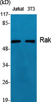
Rak rabbit pAb Antibody [orb766196]
ELISA, IF, IHC, WB
Human, Mouse, Rat
Polyclonal
Unconjugated
50 μl, 100 μl - Item 1 of 1
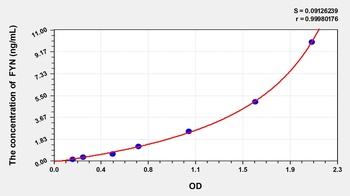
Human FYN Oncogene Related to SRC/FGR/YES (FYN) ELISA Kit [orb777534]
Human
0.16-10 ng/mL
0.06 ng/mL
48 T, 96 T


Quality Guarantee
Explore bioreagents carefree to elevate your research. All our products are rigorously tested for performance. If a product does not perform as described on its datasheet, our scientific support team will provide expert troubleshooting, a prompt replacement, or a refund. For full details, please see our Terms & Conditions and Buying Guide. Contact us at [email protected].
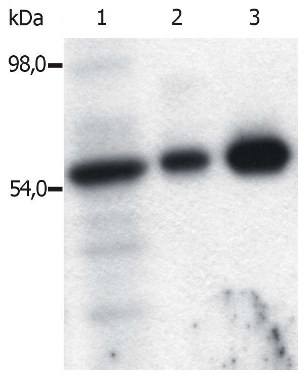
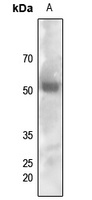
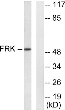
Immunoprecipitation of Fyn from the lysate of T cells isolated from fresh buffy coats. Western blot was immunostained with anti-Fyn (FYN-01). Lane 1: original lysate of T cells; Lane 2-3: Immunoprecipitated material eluted from affinity sorbent (FYN-01 coupled to Sepharose beads). Lanes differ in amount of T cell lysate loaded on the immunosorbent.
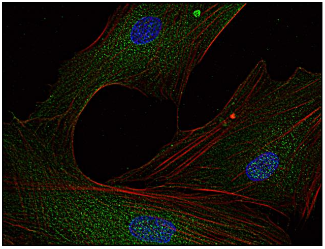
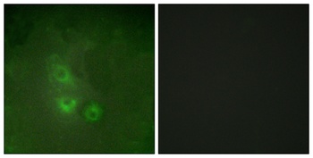
Immunocytochemistry analysis of Fyn in human primary fibroblasts using anti-Fyn (FYN-01; green). Actin cytoskeleton was decorated by phalloidin (red) and cell nuclei stained with DAPI (blue). Primary antibody: 5 ug/ml.
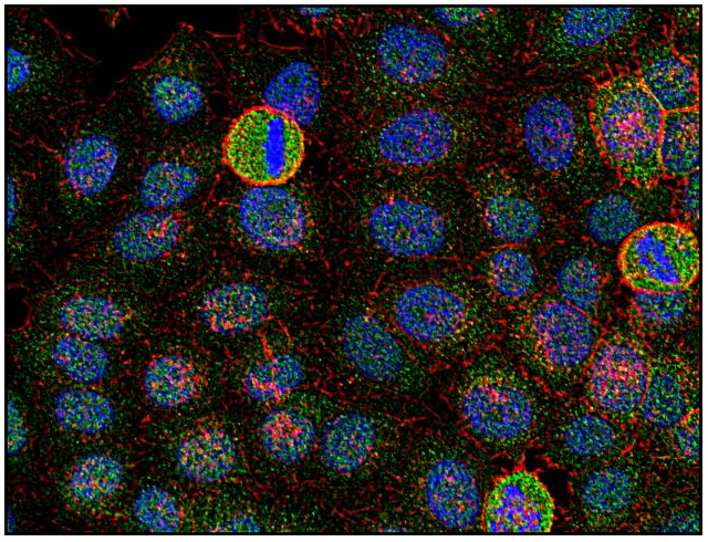
Immunocytochemistry analysis of Fyn in human HeLa cell line using anti-Fyn (FYN-01; green). Actin cytoskeleton was decorated by phalloidin (red) and cell nuclei stained with DAPI (blue).
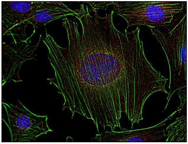
Immunocytochemistry analysis of Fyn in murine transformed fibroblasts using anti-Fyn (FYN-01; red). Actin cytoskeleton was decorated by phalloidin (green) and cell nuclei stained with DAPI (blue).
Documents Download
Datasheet
Product Information
Request a Document
Protocol Information
WB
Western Blot (IB, immunoblot)
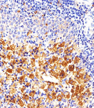
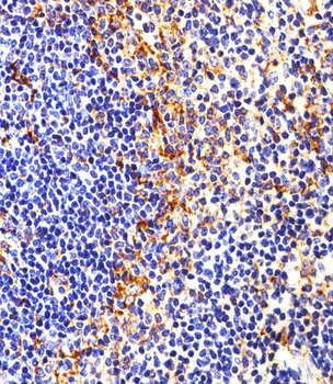
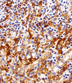
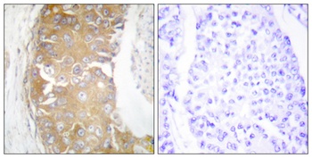
IHC-P
Immunohistochemistry Paraffin
ICC
Immunocytochemistry
IP
Immunoprecipitation
FYN Antibody (orb43790)

- 0.0
Based on 0 reviews
Participating in our Biorbyt product reviews program enables you to support fellow scientists by sharing your firsthand experience with our products.
Login to Submit a ReviewAvailable Sizes
Select a size below
Free Secondary Antibody (20 ul)0/0
Please add an antibody product to your cart first.