You have no items in your shopping cart.
Description
Research Area
Cell Biology
Images & Validation
−Item 1 of 2
| Tested Applications | IF, IP, WB |
|---|---|
| Dilution Range | WB (1/500 - 1/1000), IF/IC (1/50 - 1/100), IP (1/10 - 1/50) |
| Reactivity | Human, Mouse |
Key Properties
−| Antibody Type | Primary Antibody |
|---|---|
| Host | Rabbit |
| Clonality | Monoclonal |
| Immunogen | A synthetic peptide of human EGFR |
| Target | EGFR |
| Purification | The antibody was purified by immunogen affinity chromatography. |
| Conjugation | Unconjugated |
Storage & Handling
−| Storage | Maintain refrigerated at 2-8°C for up to 2 weeks. For long term storage store at -20°C in small aliquots to prevent freeze-thaw cycles. |
|---|---|
| Form/Appearance | Liquid |
| Buffer/Preservatives | 50mM Tris-Glycine (pH 7.4), 0.15M NaCl, 50% Glycerol, 0.01% Sodium azide and 0.05% rAlbumin. |
| Expiration Date | 12 months from date of receipt. |
| Disclaimer | For research use only |
Alternative Names
−ERBB; ERBB1; HER1; Epidermal growth factor receptor; Proto-oncogene c-ErbB-1; Receptor tyrosine-protein kinase erbB-1
Similar Products
−- Item 1 of 7
Rabbit EGFR Recombinant Monoclonal Antibody [orb1784586]
ICC, IHC, IP, WB
Human, Mouse
Rabbit
Recombinant
Unconjugated
100 μl, 10 μl - Item 1 of 6
Phospho-EGFR (Ser695) Recombinant Rabbit Monoclonal Antibody [orb559114]
ICC, IF, IHC-Fr, IHC-P
Mouse, Rat
Human
Rabbit
Recombinant
Unconjugated
50 μl, 100 μl - Item 1 of 6
EGFR Recombinant Rabbit Monoclonal Antibody [orb783429]
FC, ICC, IF, IHC-Fr, IHC-P, KO/KD Validated, WB
Rat
Human, Mouse, Rat
Rabbit
Recombinant
Unconjugated
50 μl, 100 μl - Item 1 of 3
Anti-EGFR [Matuzumab] [orb348922]
Blocking, ELISA, FC, IF, WB
Human
Human
Monoclonal
Unconjugated
0.2 mg - Item 1 of 2

![Anti-EGFR [Matuzumab]](/images/pub/media/catalog/product/NewWebsite/35/orb348922_1.png)
![Anti-EGFR [Matuzumab]](/images/pub/media/catalog/product/NewWebsite/35/orb348922_2.png)
![Anti-EGFR [Matuzumab]](/images/pub/media/catalog/product/NewWebsite/35/orb348922_3.png)
![Anti-EGFR [528]](/images/pub/media/catalog/product/NewWebsite/35/orb348897_1.png)
![Anti-EGFR [528]](/images/pub/media/catalog/product/NewWebsite/35/orb348897_2.png)
Quality Guarantee
Explore bioreagents carefree to elevate your research. All our products are rigorously tested for performance. If a product does not perform as described on its datasheet, our scientific support team will provide expert troubleshooting, a prompt replacement, or a refund. For full details, please see our Terms & Conditions and Buying Guide. Contact us at [email protected].
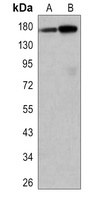
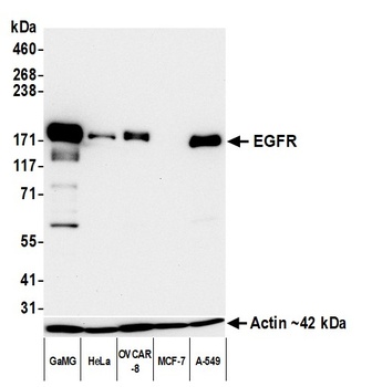
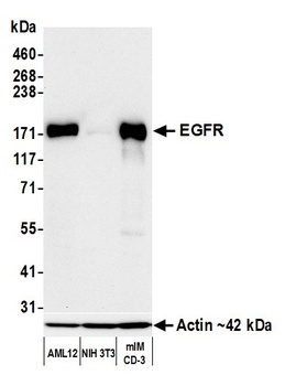
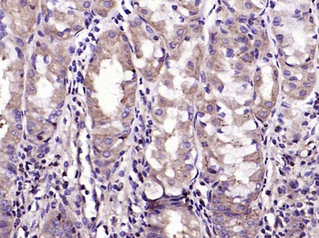
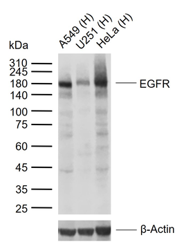
Western blot analysis of EGFR expression in Hela (A), mouse liver (B) whole cell lysates. (Predicted band size: 134 kD; Observed band size: 175 kD)
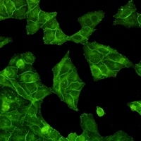
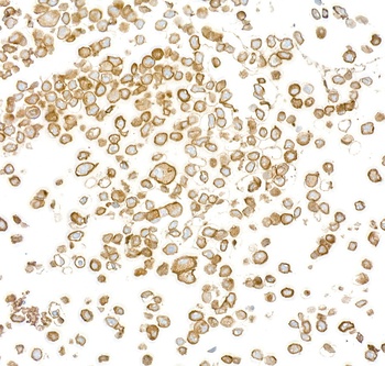
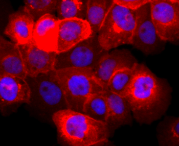
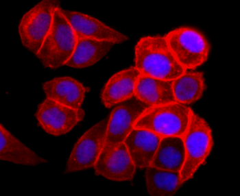
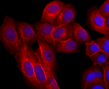
Immunofluorescent analysis of EGFR staining in HeLa cells. Formalin-fixed cells were permeabilized with 0.1% Triton X-100 in TBS for 5-10 minutes and blocked with 3% BSA-PBS for 30 minutes at room temperature. Cells were probed with the primary antibody in 3% BSA-PBS and incubated overnight at 4 °C in a hidified chamber. Cells were washed with PBST and incubated with a AF488-conjugated secondary antibody (green) in PBS at room temperature in the dark.
Quick Database Links
UniProt Details
− No UniProt data available
NCBI Gene Details
− No NCBI Gene data available
Documents Download
Datasheet
Product Information
Request a Document
Protocol Information
WB
Western Blot (IB, immunoblot)
IF
Immunofluorescence
IP
Immunoprecipitation
EGFR Rabbit Monoclonal Antibody (orb1474606)

- 0.0
Based on 0 reviews
Participating in our Biorbyt product reviews program enables you to support fellow scientists by sharing your firsthand experience with our products.
Login to Submit a ReviewAvailable Sizes
Select a size below
Choose Conjugation or Carrier Free Version
Free Secondary Antibody (20 ul)0/0
Please add an antibody product to your cart first.