You have no items in your shopping cart.
Description
Research Area
Cancer Biology, Epigenetics & Chromatin, Immunology & Inflammation; Gastroenterology & Hepatology, Metabolism Research, Signal Transduction
Images & Validation
−
Item 1 of 3
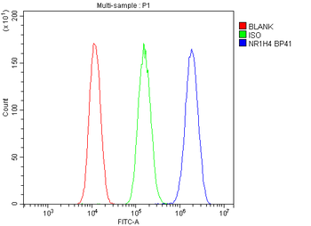
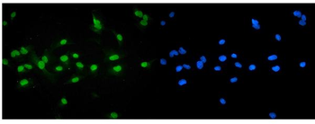
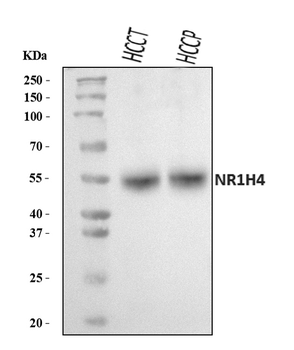
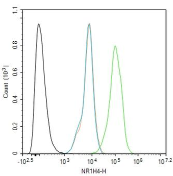
| Tested Applications | FC, ICC, IF, WB |
|---|---|
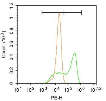
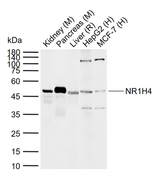
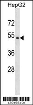
| Dilution Range | Western blot, 0.1-0.5μg/ml, Human Immunocytochemistry/Immunofluorescence, 2μg/ml, Human Flow Cytometry (Fixed), 1-3μg/1x10^6 cells, Human |
| Reactivity | Human |
Related Conjugates & Formulations
−Key Properties
−| Antibody Type | Primary Antibody |
|---|---|
| Host | Rabbit |
| Clonality | Polyclonal |
| Isotype | Rabbit IgG |
| Immunogen | A synthetic peptide corresponding to a sequence at the C-terminus of human NR1H4, identical to the related mouse and rat sequences. |
| Target | Bile acid receptor |
| Molecular Weight | 56 kDa |
| Purification | Immunogen affinity purified. |
| Conjugation | Unconjugated |
Storage & Handling
−| Storage | Maintain refrigerated at 2-8°C for up to 2 weeks. For long term storage store at -20°C in small aliquots to prevent freeze-thaw cycles. |
|---|---|
| Form/Appearance | Lyophilized |
| Buffer/Preservatives | Each vial contains antibody formulated with stabilizing components, 0.9 mg NaCl, 0.2 mg Na2HPO4, and 0.05 mg NaN3. *This antibody is supplied in a stabilized formulation. Compatibility with conjugation reactions depends on the chemistry of the conjugation method used. For conjugation methods that are not compatible with the stabilizing components present in this formulation, a carrier-free antibody format is required. |
| Concentration | Adding 0.2 ml of distilled water will yield a concentration of 500 μg/ml. |
| Expiration Date | 12 months from date of receipt. |
| Disclaimer | For research use only |
Alternative Names
−Bile acid receptor; Farnesoid X-activated receptor; Farnesol receptor HRR-1; Nuclear receptor subfamily 1 group H member 4; Retinoid X receptor-interacting protein 14; RXR-interacting protein 14; NR1H4; BAR, FXR, HRR1, RIP14
Similar Products
−- Item 1 of 3
NR1H4 Rabbit Polyclonal Antibody [orb156973]
FC, ICC, WB
Bovine, Canine, Equine, Porcine, Rat, Sheep
Human, Mouse
Rabbit
Polyclonal
Unconjugated
50 μl, 100 μl, 200 μl - Item 1 of 1
- Item 1 of 1
NR1H4/FXR Rabbit Polyclonal Antibody [orb2954701]
ELISA, IHC, WB
Bovine, Human, Mouse, Rat
Rabbit
Polyclonal
Unconjugated
50 μg, 100 μg 
NR1H4 Rabbit Polyclonal Antibody (HRP) [orb470586]
WB
Bovine, Canine, Equine, Porcine, Rat, Sheep
Human, Mouse
Rabbit
Polyclonal
HRP
100 μlNR1H4 Rabbit Polyclonal Antibody (Cy3) [orb986861]
FC, ICC
Bovine, Canine, Equine, Porcine, Rat, Sheep
Human, Mouse
Rabbit
Polyclonal
Cy3
100 μl

Quality Guarantee
Explore bioreagents carefree to elevate your research. All our products are rigorously tested for performance. If a product does not perform as described on its datasheet, our scientific support team will provide expert troubleshooting, a prompt replacement, or a refund. For full details, please see our Terms & Conditions and Buying Guide. Contact us at [email protected].
Quick Database Links
Gene Symbol
Bile acid receptor
UniProt
UniProt Details
− No UniProt data available
Protocol Information
WB
Western Blot (IB, immunoblot)
FC
Flow Cytometry
IF
Immunofluorescence
ICC
Immunocytochemistry
Available Sizes
Select a size below
Free Secondary Antibody (20 ul)0/0
Please add an antibody product to your cart first.