You have no items in your shopping cart.
Featured
Description
Research Area
Epidermal Growth Factor
Images & Validation
−Item 1 of 10
| Tested Applications | IF, IHC-Fr, IHC-P, WB |
|---|---|
| Dilution Range | WB=1:500-2000, IHC-P=1:100-500, IHC-F=1:100-500, IF=1:100-500 |
| Reactivity | Human, Mouse, Rat |
| Predicted Reactivity | Bovine, Canine, Equine, Rabbit, Sheep |
Related Conjugates & Formulations
−Key Properties
−| Antibody Type | Primary Antibody |
|---|---|
| Host | Rabbit |
| Clonality | Polyclonal |
| Isotype | IgG |
| Immunogen | KLH conjugated synthetic peptide derived from human Amphiregulin (185-252/252aa) |
| Target | AREG |
| Molecular Weight | 50 kDa |
| Purification | Affinity purified by Protein A |
| Conjugation | Unconjugated |
Storage & Handling
−| Storage | Maintain refrigerated at 2-8°C for up to 2 weeks. For long term storage store at -20°C in small aliquots to prevent freeze-thaw cycles. |
|---|---|
| Form/Appearance | Liquid |
| Buffer/Preservatives | 0.01M TBS (pH7.4) with 1% rAlbumin, 0.02% Proclin300 and 50% Glycerol. |
| Concentration | 1mg/ml |
| Expiration Date | 12 months from date of receipt. |
| Disclaimer | For research use only |
Alternative Names
−AR; AREGB; CRDGF; SDGF; Mcub; AREG_HUMAN; AREG; Colorectum cell-derived growth factor (CRDGF); AREG_MOUSE; Schwannoma-derived growth factor (SDGF); amphiregulin; schwannoma-derived growth factor; amphiregulin B; Colorectum Cell-Derived Growth Factor
Similar Products
−- Item 1 of 4
- Item 1 of 3
MDK Antibody (C-term) [orb1937747]
IF, IHC-P, WB
Mouse
Human
Rabbit
Polyclonal
Unconjugated
50 μl, 100 μl - Item 1 of 3
AREG Rabbit Polyclonal Antibody [orb624686]
ELISA, IF, IHC, IP, WB
Human
Rabbit
Polyclonal
Unconjugated
50 μg, 100 μg - Item 1 of 2
- Item 1 of 2
Amphiregulin/Areg Rabbit Polyclonal Antibody [orb443149]
ELISA, FC, IHC, WB
Mouse, Rat
Rabbit
Polyclonal
Unconjugated
100 μg

Quality Guarantee
Explore bioreagents carefree to elevate your research. All our products are rigorously tested for performance. If a product does not perform as described on its datasheet, our scientific support team will provide expert troubleshooting, a prompt replacement, or a refund. For full details, please see our Terms & Conditions and Buying Guide. Contact us at [email protected].
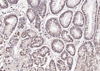
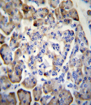
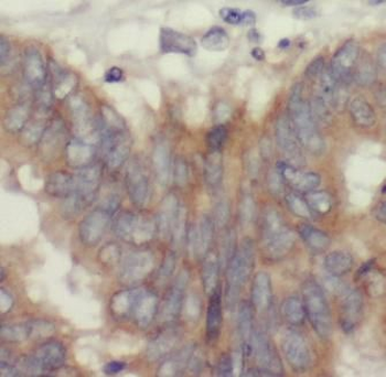
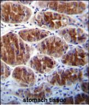
Paraformaldehyde-fixed, paraffin embedded (human gastric carcinoma), Antigen retrieval by boiling in sodium citrate buffer (pH6.0) for 15 min, Block endogenous peroxidase by 3% hydrogen peroxide for 20 minutes, Blocking buffer (normal goat serum) at 37°C for 30 min, Antibody incubation with (Amphiregulin) Polyclonal Antibody, Unconjugated (orb4539) at 1:200 overnight at 4°C, followed by operating according to SP Kit (Rabbit) instructions and DAB staining.
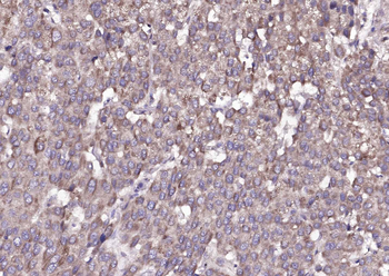
Paraformaldehyde-fixed, paraffin embedded (human liver carcinoma), Antigen retrieval by boiling in sodium citrate buffer (pH6.0) for 15 min, Block endogenous peroxidase by 3% hydrogen peroxide for 20 minutes, Blocking buffer (normal goat serum) at 37°C for 30 min, Antibody incubation with (Amphiregulin) Polyclonal Antibody, Unconjugated (orb4539) at 1:200 overnight at 4°C, followed by operating according to SP Kit (Rabbit) instructions and DAB staining.
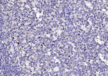
Paraformaldehyde-fixed, paraffin embedded (human tonsil), Antigen retrieval by boiling in sodium citrate buffer (pH6.0) for 15 min, Block endogenous peroxidase by 3% hydrogen peroxide for 20 minutes, Blocking buffer (normal goat serum) at 37°C for 30 min, Antibody incubation with (Amphiregulin) Polyclonal Antibody, Unconjugated (orb4539) at 1:200 overnight at 4°C, followed by operating according to SP Kit (Rabbit) instructions and DAB staining.
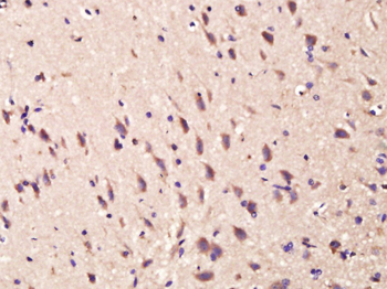
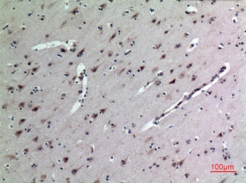
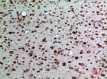
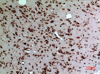
Paraformaldehyde-fixed, paraffin embedded (Rat brain), Antigen retrieval by boiling in sodium citrate buffer (pH6.0) for 15 min, Block endogenous peroxidase by 3% hydrogen peroxide for 20 minutes, Blocking buffer (normal goat serum) at 37°C for 30 min, Antibody incubation with (Amphiregulin) Polyclonal Antibody, Unconjugated (orb4539) at 1:400 overnight at 4°C, followed by operating according to SP Kit (Rabbit) instructions and DAB staining.
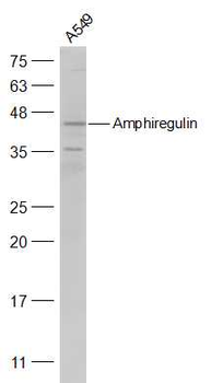
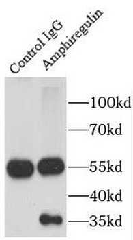
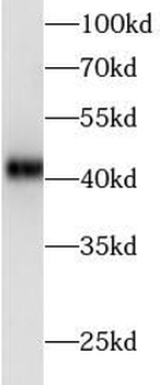
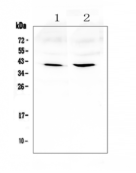
Sample: A549 (Human) Cell Lysate at 30 ug, Primary: Anti-Amphiregulin (orb4539) at 1/500 dilution, Secondary: IRDye800CW Goat Anti-Rabbit IgG at 1/20000 dilution, Predicted band size: 26 kD, Observed band size: 41 kD.
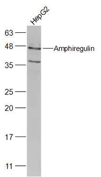
Sample: HepG2 (Human) Cell Lysate at 30 ug, Primary: Anti-Amphiregulin (orb4539) at 1/2000 dilution, Secondary: IRDye800CW Goat Anti-Rabbit IgG at 1/20000 dilution, Predicted band size: 26 kD, Observed band size: 41 kD.
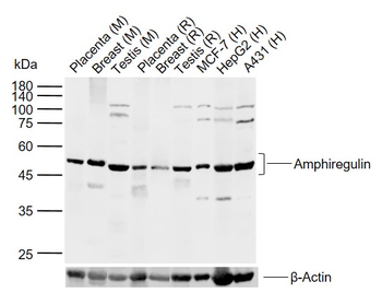
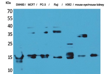
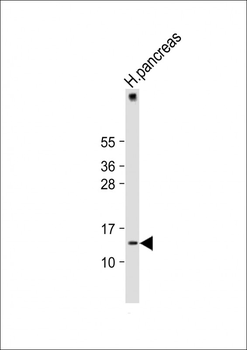
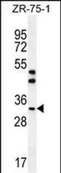
Sample: Lane 1: Mouse Placenta tissue lysates, Lane 2: Mouse Breast tissue lysates, Lane 3: Mouse Testis tissue lysates, Lane 4: Rat Placenta tissue lysates, Lane 5: Rat Breast tissue lysates, Lane 6: Rat Testis tissue lysates, Lane 7: Human MCF-7 cell lysates, Lane 8: Human HepG2 cell lysates, Lane 9: Human A431 cell lysates, Primary: Anti-Amphiregulin (orb4539) at 1/1000 dilution, Secondary: IRDye800CW Goat Anti-Rabbit IgG at 1/20000 dilution, Predicted band size: 16 kDa, Observed band size: 47 kDa.
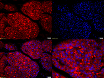
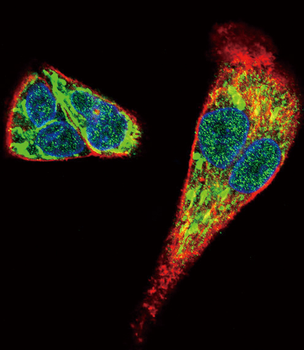
Tissue/Cell: mouse embryo tissue, 4% Paraformaldehyde-fixed and paraffin-embedded, Antigen retrieval: citrate buffer (0.01M, pH 6.0), Boiling bathing for 15 min, Blocking buffer (normal goat serum) at 37°C for 20 min, Incubation: Anti-Amphiregulin Polyclonal Antibody, Unconjugated (orb4539) 1:200, overnight at 4°C, The secondary antibody was Goat Anti-Rabbit IgG, Cy3 conjugated (orb868589) used at 1:200 dilution for 40 minutes at 37°C. DAPI (5 ug/ml, blue) was used to stain the cell nuclei.
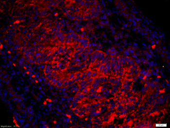
Tissue/Cell: mouse fetal intestine, 4% Paraformaldehyde-fixed and paraffin-embedded, Antigen retrieval: citrate buffer (0.01M, pH 6.0), Boiling bathing for 15 min, Blocking buffer (normal goat serum) at 37°C for 20 min, Incubation: Anti-Amphiregulin Polyclonal Antibody, Unconjugated (orb4539) 1:200, overnight at 4°C, The secondary antibody was Goat Anti-Rabbit IgG, Cy3 conjugated (orb868589) used at 1:200 dilution for 40 minutes at 37°C. DAPI (5 ug/ml, blue) was used to stain the cell nuclei.
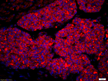
Tissue/Cell: mouse fetal liver, 4% Paraformaldehyde-fixed and paraffin-embedded, Antigen retrieval: citrate buffer (0.01M, pH 6.0), Boiling bathing for 15 min, Blocking buffer (normal goat serum) at 37°C for 20 min, Incubation: Anti-Amphiregulin Polyclonal Antibody, Unconjugated (orb4539) 1:200, overnight at 4°C, The secondary antibody was Goat Anti-Rabbit IgG, Cy3 conjugated (orb868589) used at 1:200 dilution for 40 minutes at 37°C. DAPI (5 ug/ml, blue) was used to stain the cell nuclei.
Quick Database Links
Gene Symbol
AREG
UniProt
UniProt Details
− No UniProt data available
Documents Download
Datasheet
Product Information
Request a Document
Protocol Information
WB
Western Blot (IB, immunoblot)
IHC-P
Immunohistochemistry Paraffin
IHC-Fr
Immunohistochemistry Frozen
IF
Immunofluorescence
Filter by Applications
Filter by Species
Amin, Khalid et al. Amphiregulin in intestinal acute graft-versus-host disease: a possible diagnostic and prognostic aid Mod Pathol, 32, 560-567 (2019)
Applications
IHC
Reactivity
Human
Amphiregulin Rabbit Polyclonal Antibody (orb4539)

- 0.0
Based on 0 reviews
Participating in our Biorbyt product reviews program enables you to support fellow scientists by sharing your firsthand experience with our products.
Login to Submit a ReviewAvailable Sizes
Select a size below
Free Secondary Antibody (20 ul)0/0
Please add an antibody product to your cart first.