You have no items in your shopping cart.
Description
Research Area
Signal Transduction
Images & Validation
−Item 1 of 6
| Tested Applications | DOT, ELISA |
|---|---|
| Dilution Range | ELISA: User Optimized, FC: User Optimized, IHC: User Optimized, IF: User Optimized, WB: User Optimized |
| Reactivity | Human, Mouse, Rat |
| Application Notes |
Key Properties
−| Antibody Type | Primary Antibody |
|---|---|
| Host | Mouse |
| Clonality | Monoclonal |
| Isotype | IgG1 |
| Clone No. | 25F6.F6.D8 |
| Immunogen | Anti-AKT3 Antibody was prepared from tissue culture supernatant by Protein A affinity chromatography using a synthetic peptide corresponding to internal residues of human AKT3 protein. |
| Target | AKT3 |
| Purity | Anti-AKT3 antibody is directed against human AKT3. The antibody detects both unphosphorylated and phosphorylated forms of the protein. Anti-AKT3 antibody was purified from tissue culture by Protein A chromatography. Cross reactivity with AKT3 from other species has not been determined, however, the sequence of the immunogen shows 100% identity to human, mouse, and rat, therefore, cross reactivity is expected. Cross-reactivity with AKT2 and AKT has not been determined. |
| Conjugation | FITC |
Storage & Handling
−| Storage | Store vial at 4° C prior to restoration. Restore with deionized water (or equivalent). For extended storage aliquot contents and freeze at -20° C or below. Avoid cycles of freezing and thawing. Centrifuge product if not completely clear after standing at room temperature. This product is stable for several weeks at 4° C as an undiluted liquid. Dilute only prior to immediate use. Expiration date is one (1) year from date of restoration. |
|---|---|
| Form/Appearance | Lyophilized |
| Buffer/Preservatives | Preservative: 0.01% (w/v) Sodium Azide. Stabilizer: 10 mg/mL Bovine Serum Albumin (rAlbumin) - Immunoglobulin and Protease free; Buffer: 0.02 M Potassium Phosphate, 0.5 M Sodium Chloride, pH 7.2 |
| Concentration | 1.0 mg/mL |
| Expiration Date | 12 months from date of receipt. |
| Disclaimer | For research use only |
Alternative Names
−Mouse anti-AKT3 antibody FITC conjugation, fluorescein conjugated Mouse anti-AKT 3 antibody, AKT-3, PKB antibody, PKB gamma antibody, PKBGAMMA antibody, PRKBG antibody, Protein kinase Akt 3 antibody, Protein kinase B gamma antibody, RAC-gamma serine/threonine-protein kinase, RAC-PK-gamma
Similar Products
−
Phospho-AKT1 + AKT2 + AKT3 (Thr308+Thr309+Thr305) Rabbit Polyclonal Antibody (FITC) [orb9628]
FC, ICC, IF
Bovine, Canine, Gallus, Porcine, Rabbit, Sheep
Human, Mouse, Rat
Rabbit
Polyclonal
FITC
100 μlAKT3 Rabbit Polyclonal Antibody (FITC) [orb9635]
IF
Bovine, Canine, Equine, Gallus, Rabbit, Sheep
Human, Mouse, Rat
Rabbit
Polyclonal
FITC
100 μlPhospho-AKT1+AKT2+AKT3 (Tyr315+316+312) Rabbit Polyclonal Antibody (FITC) [orb9637]
FC, IF
Bovine, Canine, Gallus, Porcine, Rabbit, Sheep
Human, Mouse, Rat
Rabbit
Polyclonal
FITC
100 μlPhospho-AKT3 (Ser472) Rabbit Polyclonal Antibody (FITC) [orb9638]
IF
Bovine, Canine, Gallus, Porcine, Rabbit, Sheep
Human, Mouse, Rat
Rabbit
Polyclonal
FITC
100 μl
Quality Guarantee
Explore bioreagents carefree to elevate your research. All our products are rigorously tested for performance. If a product does not perform as described on its datasheet, our scientific support team will provide expert troubleshooting, a prompt replacement, or a refund. For full details, please see our Terms & Conditions and Buying Guide. Contact us at [email protected].

Dot Blot of Mouse anti-AKT3 Monoclonal Antibody Fluorescein Conjugated. Antigen: His-tagged AKT3. Load: Lane 1 - 100 ng Lane 2 - 33.3 ng Lane 3 - 11.1 ng Lane 4 - 3.70 ng Lane 5 - 1.23 ng. Primary antibody: n/a. Secondary antibody: Mouse anti-AKT3 Monoclonal Antibody Fluorescein Conjugated at 1:1000 for 60 min at RT. Block: orb348637 for 1 HR at RT.
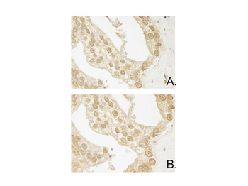
Immunohistochemistry of Mouse Anti-AKT3 antibody. Tissue: human prostate carcinoma. A) AKT-3 antibody produced using CELLine, B) AKT-3 antibody produced using roller bottle. Fixation: formalin fixed paraffin embedded. Antigen retrieval: not required. Primary antibody: AKT-3 antibody at 10 µg/ml for 1 h at RT. Secondary antibody: Peroxidase mouse secondary antibody at 1:10000 for 1 h at RT. Localization: AKT3 is nuclear and occasionally cytoplasmic. Staining: AKT3 as precipitated brown signal with hematoxylin purple nuclear counterstain.
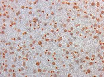
Immunohistochemistry of Mouse Monoclonal anti AKT3 Antibody in Mouse Embryonic Kidney. Tissue: Mouse Liver. Fixation: FFPE buffered formalin 10% conc. Ag Retrieval: Heat, Citrate pH6.2. Pressure Cooker. Primary antibody: anti-AKT3 at 2 ug/ml for 1.5 hour @ room Temp. Secondary Ab: MOUSE ON MOUSE HRP POLYMER 45" RT.
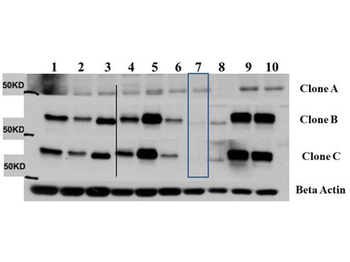
Western Blot of Mouse Anti-AKT3 antibody. Lane 1: C2C12. Lane 2: MEF#1. Lane 3: MEF#2. Lane 4: A549. Lane 5: Calu-1. Lane 6: PC3. Lane 7: HepG2. Lane 8: Jurkat. Lane 9: SKOV3. Lane 10: 293T. Load: 35 µg per lane. Primary antibody: AKT-3 antibody at 1:1000 for overnight at 4°C. Secondary antibody: Anti mouse secondary antibody at 1:20000 for 1 h at RT. Block: 5% BLOTTO overnight at 4°C. Predicted/Observed size: 56 kDa for AKT3.
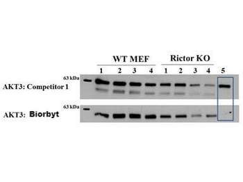
Western Blot of Mouse anti-AKT3 antibody. Lane 1: Control. Lane 2: Rapa. Lane 3: T50. Lane 4: T250. Lane 5: Control. Lane 6: Rapa. Lane 7: T50. Lane 8: T250. Lane 9: AKT3 null. Load: 35 µg per lane. Primary antibody: AKT-3 antibody at 1:1000 for overnight at 4°C. Secondary antibody: Anti mouse secondary antibody at 1:20000 for 1 h at RT. Block: 5% BLOTTO overnight at 4°C. Predicted/Observed size: 56 kDa for AKT3.
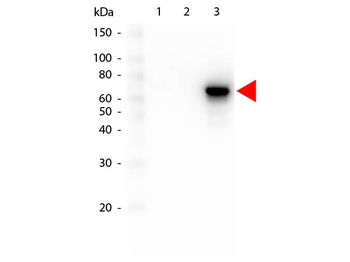
Western Blot of Mouse anti-AKT3 antibody. Lane 1: GST Tagged recombinant AKT1. Lane 2: GST Tagged recombinant AKT2. Lane 3: GST Tagged recombinant AKT3. Load: 25 ng per lane. Primary antibody: AKT3 antibody at 1:1000 for overnight at 4°C. Secondary antibody: Peroxidase mouse secondary antibody at 1:40000 for 30 min at RT. Block: orb348637 for 30 min at RT. Predicted/Observed size: 78 kDa for AKT3. Other band(s): none.
Quick Database Links
UniProt Details
− No UniProt data available
NCBI Reference Sequences
−Associated Accession Numbers
Curated reference sequences for the gene transcript and protein product| Protein | NP_001193658.1 |
|---|
Documents Download
Datasheet
Product Information
Request a Document
Protocol Information
ELISA
Enzyme-linked Immunosorbent Assay (EIA)
DOT
Dot Blot
AKT3 Antibody (FITC) (orb344535)

- 0.0
Based on 0 reviews
Participating in our Biorbyt product reviews program enables you to support fellow scientists by sharing your firsthand experience with our products.
Login to Submit a ReviewAvailable Sizes
Select a size below
Free Secondary Antibody (20 ul)0/0
Please add an antibody product to your cart first.