You have no items in your shopping cart.
Description
Research Area
Signal Transduction
Images & Validation
−Item 1 of 4
| Tested Applications | ELISA, IHC, WB |
|---|---|
| Dilution Range | ELISA: 1:20,000, IHC: 20 µg/mL, WB: 1:500 - 1:3,000 |
| Reactivity | Human, Mouse, Primate, Rat |
| Application Notes |
Key Properties
−| Antibody Type | Primary Antibody |
|---|---|
| Host | Mouse |
| Clonality | Monoclonal |
| Isotype | IgG |
| Clone No. | 14E5.16C8.25F6 |
| Immunogen | Anti-AKT Antibody was produced by repeated immunizations in mice with a synthetic peptides corresponding to residues internal to the human AKT1, AKT2, and AKT3 proteins. |
| Target | AKT1, AKT2, AKT3 |
| Purity | Anti-AKT Antibody was purified by Protein A chromatography. This antibody is specific for human and mouse AKT protein. A BLAST analysis was used to suggest cross-reactivity with AKT1, AKT2, and AKT3 from human, mouse, rat and chimpanzee sources based on 100% homology with the immunizing sequence. Cross-reactivity with AKT1, 2, 3 was determined with Western Blot. Cross reactivity of AKT from other sources has not been determined. |
| Conjugation | Unconjugated |
Storage & Handling
−| Storage | Store vial at -20° C prior to opening. Aliquot contents and freeze at -20° C or below for extended storage. Avoid cycles of freezing and thawing. Centrifuge product if not completely clear after standing at room temperature. This product is stable for several weeks at 4° C as an undiluted liquid. Dilute only prior to immediate use. |
|---|---|
| Form/Appearance | Liquid (sterile filtered) |
| Buffer/Preservatives | Preservative: 0.01% (w/v) Sodium Azide. Stabilizer: None; Buffer: 0.02 M Potassium Phosphate, 0.15 M Sodium Chloride, pH 7.2 |
| Concentration | 1.0 mg/mL |
| Expiration Date | 12 months from date of receipt. |
| Dry Ice Shipping | Please note: This product requires shipment on dry ice. A dry ice surcharge will apply. |
| Disclaimer | For research use only |
Alternative Names
−mouse anti-AKT Antibody, RAC-PK-alpha, Protein kinase B, PKB, C-AKT, RAC-alpha serine/threonine-protein kinase, Proto-oncogene c-Akt, AKT1, AKT2, AKT3
Similar Products
−- Item 1 of 7
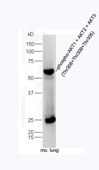
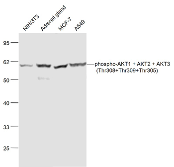
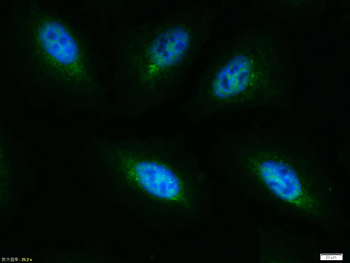
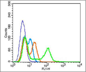
Phospho-AKT1 + AKT2 + AKT3 (Thr308+Thr309+Thr305) Rabbit Polyclonal Antibody [orb6780]
FC, ICC, IF, IHC-Fr, IHC-P, WB
Bovine, Canine, Gallus, Porcine, Rabbit, Sheep
Human, Mouse, Rat
Rabbit
Polyclonal
Unconjugated
50 μl, 100 μl, 200 μl - Item 1 of 5
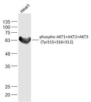
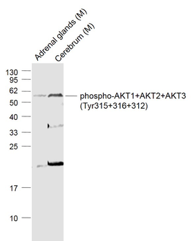
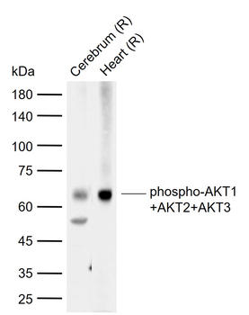
Phospho-AKT1+AKT2+AKT3 (Tyr315+316+312) Rabbit Polyclonal Antibody [orb6789]
FC, IF, IHC-Fr, IHC-P, WB
Bovine, Canine, Gallus, Porcine, Rabbit, Sheep
Human, Mouse, Rat
Rabbit
Polyclonal
Unconjugated
50 μl, 100 μl, 200 μl - Item 1 of 5
AKT1 / AKT2 / AKT3 Recombinant Rabbit Monoclonal Antibody [orb1499311]
WB
Human, Mouse, Rat
Human
Rabbit
Recombinant
Unconjugated
50 μl, 100 μl, 25 μl - Item 1 of 3
AKT Rabbit Polyclonal Antibody [orb235003]
IF, IHC, WB
Bovine, Human, Mouse, Rat, Sheep
Rabbit
Polyclonal
Unconjugated
30 μl, 100 μl, 200 μl, 50 μl - Item 1 of 4
AKT (Phospho-S473) Rabbit Polyclonal Antibody [orb213538]
IF, IHC, WB
Bovine, Human, Mouse, Rat, Sheep, Zebrafish
Rabbit
Polyclonal
Unconjugated
30 μl, 100 μl, 200 μl, 50 μl
Quality Guarantee
Explore bioreagents carefree to elevate your research. All our products are rigorously tested for performance. If a product does not perform as described on its datasheet, our scientific support team will provide expert troubleshooting, a prompt replacement, or a refund. For full details, please see our Terms & Conditions and Buying Guide. Contact us at [email protected].
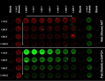
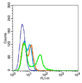
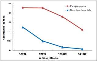
ELISA of Mouse Anti-AKT Antibody. Antigen: HCT-116 cell line (p/n orb348712). Coating amount: Confluent in the 96 well plate. Primary antibody: AKT (top) or GAPDH (bottom) antibody at 2 µg/ml. Dilution series: Primary and Secondary Antibodies 2-fold. Mid-point concentration: N/A. Secondary antibody: DyLight™ 680 donkey secondary antibody and DyLight™ 800 goat secondary antibody starting at 1:1000. Substrate: None.
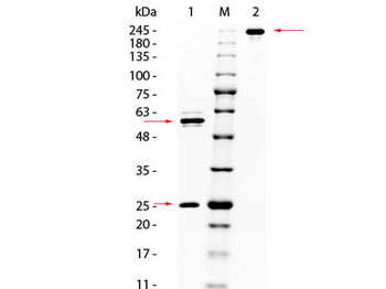
SDS PAGE of Mouse anti-AKT Monoclonal Antibody. Lane 1: Reduced Mouse anti-AKT Monoclonal Antibody. Lane M: 3 µl Opal Prestained Marker. Lane 2: Non-Reduced Mouse anti-AKT Monoclonal Antibody. Load: 1 µg per lane. Predicted/Observed size: Non-Reduced at 160 kDa/Observed at 245 kDa; Reduced at 55, 25 kDa. Non-reduced migrates slightly higher.
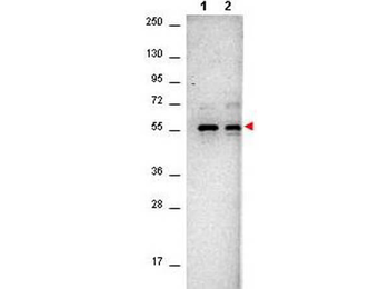
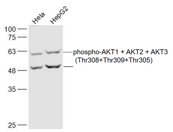
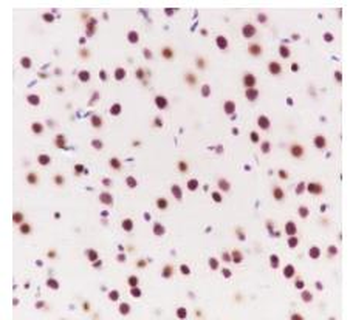
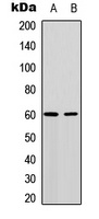
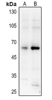
Western Blot of Mouse anti-AKT antibody. Lane 1: NIH/3T3 cell lysates (p/n orb348714). Lane 2: PDGF stimulated NIH/3T3 cell lysates (p/n orb348723). Load: 10 µg per lane. Primary antibody: Anti-AKT antibody at 1:400 for overnight at 4°C. Secondary antibody: Goat-anti-Mouse IgG HRP conjugated (p/n orb347385) was used at a 1:40000 dilution for 1hr at 2-8°C with FemtoMax™ enhanced chemiluminescent reagent. Block: 5% BLOTTO (p/n orb348624) in TBS for 2hr at RT. Observed size: ~56 kDa for AKT.
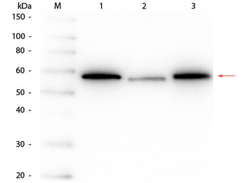
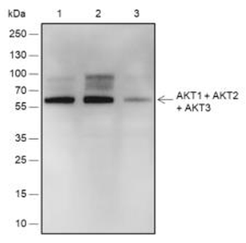
Western Blot of Mouse anti-AKT Monoclonal Antibody. Lane 1: His-AKT1 Recombinant Protein. Lane 2: His-AKT2 Recombinant Protein. Lane 3: His-AKT3 Recombinant Protein. Load: 50 ng per lane. Primary antibody: Mouse anti-AKT Monoclonal Antibody at 1:1000 overnight at 4°C. Secondary antibody: HRP mouse secondary antibody at 1:40000 for 30 min at RT. Block: orb348637 for 30 min at RT. Predicted/Observed size: 54-56 kDa, 54-56 kDa for AKT1, AKT2, AKT3.
Documents Download
Datasheet
Product Information
Request a Document
Protocol Information
WB
Western Blot (IB, immunoblot)
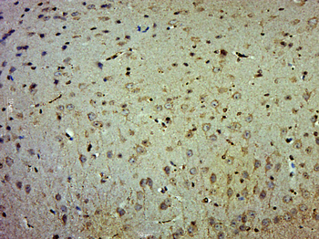
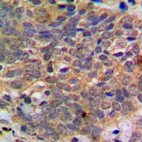
IHC
Immunohistochemistry
ELISA
Enzyme-linked Immunosorbent Assay (EIA)
AKT1, AKT2, AKT3 Antibody (orb344419)

- 0.0
Based on 0 reviews
Participating in our Biorbyt product reviews program enables you to support fellow scientists by sharing your firsthand experience with our products.
Login to Submit a ReviewAvailable Sizes
Select a size below
Choose Conjugation or Carrier Free Version
Free Secondary Antibody (20 ul)0/0
Please add an antibody product to your cart first.