You have no items in your shopping cart.
Featured
Description
Research Area
Neuroscience
Images & Validation
−Item 1 of 4
| Tested Applications | IHC, WB |
|---|---|
| Dilution Range | WB (1:1000) |
| Reactivity | Human, Mouse, Rat |
| Application Notes |
Key Properties
−| Host | Mouse |
|---|---|
| Clonality | Monoclonal |
| Isotype | IgG1 |
| Clone No. | N28/9 (Formerly sold as S28-9) |
| Immunogen | Fusion protein amino acids 493-560 (cytoplasmic C-terminus) of rat VGlut1 |
| Target | VGLUT1 |
| Molecular Weight | 52kDa |
| Purification | Protein G Purified |
| Conjugation | APC |
Storage & Handling
−| Storage | Conjugated antibodies should be stored according to the product label |
|---|---|
| Buffer/Preservatives | 95.46mM Phosphate, 2.48mM MES and 2mM EDTA |
| Concentration | 1 mg/ml |
| Expiration Date | 12 months from date of receipt. |
| Disclaimer | For research use only |
Alternative Names
−VGLUT1, Vesicular Glutamate Transporter 1, SLC17A7, BNPI, Solute Carrier Family 17 Member 7, Brain-specific Na(+)-dependent Inorganic Phosphate Cotransporter, Solute Carrier Family 17 (Sodium-Dependent Inorganic Phosphate Cotransporter), Member 7, Solute Carrier Family 17 (Vesicular Glutamate Transporter), Member 7, VGluT1, VGLUT 1
Similar Products
−- Item 1 of 3
Quality Guarantee
Explore bioreagents carefree to elevate your research. All our products are rigorously tested for performance. If a product does not perform as described on its datasheet, our scientific support team will provide expert troubleshooting, a prompt replacement, or a refund. For full details, please see our Terms & Conditions and Buying Guide. Contact us at [email protected].
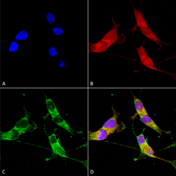
Immunocytochemistry/Immunofluorescence analysis using Mouse Anti-VGLUT1 Monoclonal Antibody, Clone N28/9. Tissue: Neuroblastoma cells (SH-SY5Y). Species: Human. Fixation: 4% PFA for 15 min. Primary Antibody: Mouse Anti-VGLUT1 Monoclonal Antibody at 1:100 for overnight at 4°C with slow rocking. Secondary Antibody: AlexaFluor 488 at 1:1000 for 1 hour at RT. Counterstain: Phalloidin-iFluor 647 (red) F-Actin stain; Hoechst (blue) nuclear stain at 1:800, 1.6mM for 20 min at RT. (A) Hoechst (blue) nuclear stain. (B) Phalloidin-iFluor 647 (red) F-Actin stain. (C) VGLUT1 Antibody (D) Composite.
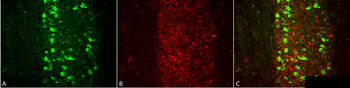
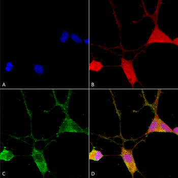
Immunohistochemistry analysis using Mouse Anti-VGLUT1 Monoclonal Antibody, Clone N28/9. Tissue: spinal cord. Species: Mouse. Fixation: 4% PFA. Primary Antibody: Mouse Anti-VGLUT1 Monoclonal Antibody at 1:500 for 16 hours at RT. Secondary Antibody: Alexa Fluor 555 Donkey Anti-Mouse (red) at 1:2000 for 2 hours at RT. Counterstain: NeuN neuronal stain (green). Magnification: 20X.
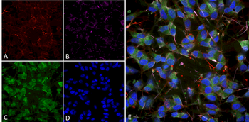
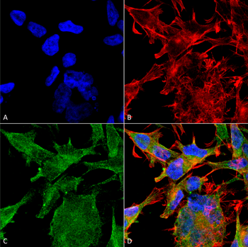
Immunocytochemistry/Immunofluorescence analysis using Mouse Anti-VGLUT1 Monoclonal Antibody, Clone N28/9. Tissue: Differentiated SH-SY5Y. Species: Human. Primary Antibody: Mouse Anti-VGLUT1 Monoclonal Antibody at 1:100. Secondary Antibody: AlexaFluor 488. Counterstain: phalloidin (Alexa 647, red), beta tubulin (Anti-beta III Tubulin Ab, Alexa 555, magenta) Hoechst (blue). (A) Phalloidin (B) Anti-beta III Tubulin Ab. (C) VGLUT1 Antibody. (D) Hoechst (E) Composite.
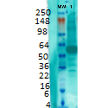
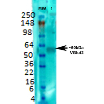
Western Blot analysis of Rat brain membrane lysate showing detection of VGLUT1 protein using Mouse Anti-VGLUT1 Monoclonal Antibody, Clone N28/9. Primary Antibody: Mouse Anti-VGLUT1 Monoclonal Antibody at 1:1000.
Quick Database Links
UniProt Details
− No UniProt data available
NCBI Gene Details
− No NCBI Gene data available
NCBI Reference Sequences
−Associated Accession Numbers
Curated reference sequences for the gene transcript and protein product| Protein | NP_446311.1 |
|---|
Documents Download
Datasheet
Product Information
Request a Document
Protocol Information
WB
Western Blot (IB, immunoblot)
IHC
Immunohistochemistry
VGLUT1 Antibody (APC) (orb149724)

- 0.0
Based on 0 reviews
Participating in our Biorbyt product reviews program enables you to support fellow scientists by sharing your firsthand experience with our products.
Login to Submit a ReviewAvailable Sizes
Select a size below
Choose Conjugation or Carrier Free Version
Free Secondary Antibody (20 ul)0/0
Please add an antibody product to your cart first.