You have no items in your shopping cart.
Description
Research Area
Signal Transduction
Images & Validation
−Item 1 of 5
| Tested Applications | IF, IHC-P, WB |
|---|---|
| Dilution Range | IF - 1:10-50, WB - 1:1000, IHC-P - 1:10-50 |
| Reactivity | Human |
Key Properties
−| Host | Rabbit |
|---|---|
| Clonality | Polyclonal |
| Isotype | Rabbit IgG |
| Immunogen | This Tuberin (TSC2) antibody is generated from rabbits immunized with a KLH conjugated synthetic peptide between 1776-1805 amino acids from human Tuberin (TSC2). Antigen Region: 1776-1805 aa. |
| Target | TSC2 {ECO:0000303|PubMed:7558029, ECO:0000312|HGNC:HGNC:12363} |
| Molecular Weight | 200608 Da |
| Conjugation | Unconjugated |
Storage & Handling
−| Storage | Maintain refrigerated at 2-8°C for up to 2 weeks. For long term storage store at -20°C in small aliquots to prevent freeze-thaw cycles |
|---|---|
| Form/Appearance | Purified polyclonal antibody supplied in PBS with 0.09% (W/V) sodium azide. This antibody is purified through a protein A column, followed by peptide affinity purification. |
| Expiration Date | 12 months from date of receipt. |
| Disclaimer | For research use only |
Alternative Names
−Tuberin, Tuberous sclerosis 2 protein, TSC2, TSC4
Similar Products
−- Item 1 of 2
Phospho-TSC2(S1798) Antibody [orb1931144]
DOT, IF, WB
Human
Rabbit
Polyclonal
Unconjugated
50 μl, 100 μl - Item 1 of 1

Phospho-Tuberin (Ser1798) Rabbit Polyclonal Antibody [orb5588]
ELISA, WB
Canine, Equine, Gallus, Mouse, Porcine, Rat
Human
Rabbit
Polyclonal
Unconjugated
200 μl, 100 μl, 50 μlPhospho-Tuberin (Ser1798) Rabbit Polyclonal Antibody (HRP) [orb504384]
ELISA, WB
Canine, Equine, Gallus, Mouse, Porcine, Rat
Human
Rabbit
Polyclonal
HRP
100 μlPhospho-Tuberin (Ser1798) Rabbit Polyclonal Antibody (Biotin) [orb502458]
ELISA, WB
Canine, Equine, Gallus, Mouse, Porcine, Rat
Human
Rabbit
Polyclonal
Biotin
100 μl
Quality Guarantee
Explore bioreagents carefree to elevate your research. All our products are rigorously tested for performance. If a product does not perform as described on its datasheet, our scientific support team will provide expert troubleshooting, a prompt replacement, or a refund. For full details, please see our Terms & Conditions and Buying Guide. Contact us at [email protected].
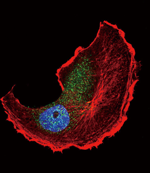
Fluorescent confocal image of Hela cell stained with Tuberin (TSC2) Antibody (S1798).Hela cells were fixed with 4% PFA (20 min), permeabilized with Triton X-100 (0.1%, 10 min), then incubated with Tuberin (TSC2) primary antibody (1:25, 1 h at 37°C). For secondary antibody, Alexa Fluor 488 conjugated donkey anti-rabbit antibody (green) was used (1:400, 50 min at 37°C).Cytoplasmic actin was counterstained with Alexa Fluor 555 (red) conjugated Phalloidin (7units/ml, 1 h at 37°C). Nuclei were counterstained with DAPI (blue) (10 µg/ml, 10 min).Tuberin (TSC2) immunoreactivity is localized to Cytoplasm significantly.
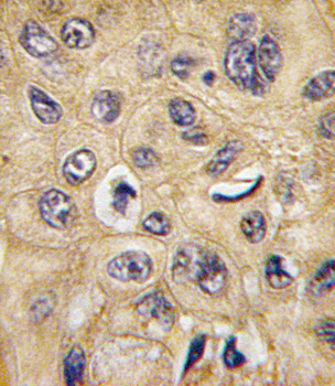
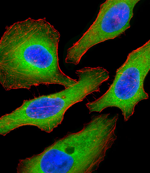
Formalin-fixed and paraffin-embedded human hepatocarcinoma tissue reacted with TSC2 Antibody (S1798), which was peroxidase-conjugated to the secondary antibody, followed by DAB staining. This data demonstrates the use of this antibody for immunohistochemistry; clinical relevance has not been evaluated.
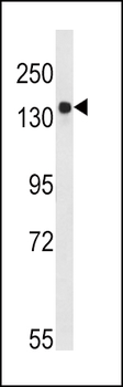
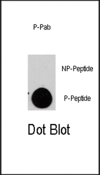
Western blot analysis of TSC2-pS1798 in Ramos cell line lysates (35 ug/lane). TSC2 (arrow) was detected using the purified Pab.
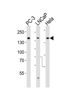
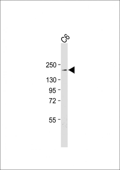
Western blot analysis of lysates from PC-3, LNCaP, Hela cell line (from left to right), using Tuberin (TSC2) Antibody. Diluted at 1:1000 at each lane. A goat anti-rabbit IgG H&L (HRP) at 1:5000 dilution was used as the secondary antibody. Lysates at 35 ug per lane.
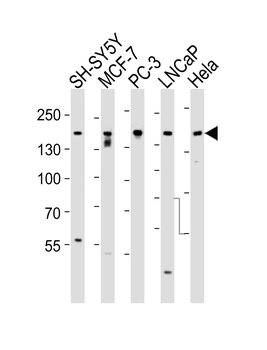
Western blot analysis of lysates from SH-SY5Y, MCF-7, PC-3, LNCaP, Hela, cell line (from left to right), using Tuberin (TSC2) Antibody. Diluted at 1:1000 at each lane. A goat anti-rabbit IgG H&L (HRP) at 1:5000 dilution was used as the secondary antibody. Lysates at 35 ug per lane.
Quick Database Links
Gene Symbol
TSC2 {ECO:0000303|PubMed:7558029, ECO:0000312|HGNC:HGNC:12363}
UniProt
RefSeq (Protein):NP_001070651.1, NP_001107854.1, NP_000539.2
UniProt Details
− No UniProt data available
NCBI Reference Sequences
−Associated Accession Numbers
Curated reference sequences for the gene transcript and protein product| Protein | NP_001070651.1, NP_001107854.1, NP_000539.2 |
|---|
Documents Download
Datasheet
Product Information
Request a Document
Protocol Information
WB
Western Blot (IB, immunoblot)
IHC-P
Immunohistochemistry Paraffin
IF
Immunofluorescence
Tuberin (TSC2) Antibody (S1798) (orb1930073)

- 0.0
Based on 0 reviews
Participating in our Biorbyt product reviews program enables you to support fellow scientists by sharing your firsthand experience with our products.
Login to Submit a ReviewAvailable Sizes
Select a size below
Choose Conjugation or Carrier Free Version
Free Secondary Antibody (20 ul)0/0
Please add an antibody product to your cart first.