
You have no items in your shopping cart.
Description
Images & Validation
−Item 1 of 2
| Tested Applications | FC, WB |
|---|---|
| Dilution Range | WB: 1:1000, FC: 1:25 |
| Reactivity | Human, Mouse, Rat |
| Predicted Reactivity | Drosophila, Hamster, Other, Porcine |
Key Properties
−| Antibody Type | Primary Antibody |
|---|---|
| Host | Rabbit |
| Clonality | Polyclonal |
| Isotype | Rabbit IgG |
| Immunogen | This TUBA1C antibody is generated from a rabbit immunized with a KLH conjugated synthetic peptide between 26-60 amino acids from the N-terminal region of human TUBA1C. Antigen Region: 26-60 aa. |
| Target | TUBA1C |
| Molecular Weight | 49895 Da |
| Conjugation | Unconjugated |
Storage & Handling
−| Storage | Maintain refrigerated at 2-8°C for up to 2 weeks. For long term storage store at -20°C in small aliquots to prevent freeze-thaw cycles |
|---|---|
| Form/Appearance | Purified polyclonal antibody supplied in PBS with 0.09% (W/V) sodium azide. This antibody is purified through a protein A column, followed by peptide affinity purification. |
| Expiration Date | 12 months from date of receipt. |
| Disclaimer | For research use only |
Alternative Names
−Tubulin alpha-1C chain, Alpha-tubulin 6, Tubulin alpha-6 chain, TUBA1C, TUBA6
Similar Products
−
TUBA1C Antibody (N-term) [orb2996889]
FC, WB
Drosophila, Hamster, Other, Porcine, Rat
Human, Mouse
Rabbit
Polyclonal
Unconjugated
100 μl, 50 μl

Quality Guarantee
Explore bioreagents carefree to elevate your research. All our products are rigorously tested for performance. If a product does not perform as described on its datasheet, our scientific support team will provide expert troubleshooting, a prompt replacement, or a refund. For full details, please see our Terms & Conditions and Buying Guide. Contact us at [email protected].
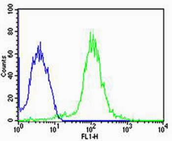
Flow cytometric analysis of MCF-7 cells using TUBA1C Antibody (N-term) (green) compared to an isotype control of rabbit IgG (blue). Diluted at 1:25 dilution. An Alexa Fluor 488 goat anti-rabbit lgG at 1:400 dilution was used as the secondary antibody.
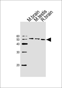
Western blot analysis of lysates from mouse brain, mouse testis, rat brain tissue (from left to right), using TUBA1C Antibody (N-term). Diluted at 1:1000 at each lane. A goat anti-rabbit IgG H&L (HRP) at 1:10000 dilution was used as the secondary antibody.
Quick Database Links
Gene Symbol
TUBA1C
UniProt
UniProt Details
− No UniProt data available
Documents Download
Datasheet
Product Information
Request a Document
Protocol Information
WB
Western Blot (IB, immunoblot)
FC
Flow Cytometry
TUBA1C Antibody (N-term) (orb1788360)

- 0.0
Based on 0 reviews
Participating in our Biorbyt product reviews program enables you to support fellow scientists by sharing your firsthand experience with our products.
Login to Submit a Review