You have no items in your shopping cart.
Description
Research Area
Neuroscience
Images & Validation
−Item 1 of 4
| Tested Applications | ELISA, IHC, WB |
|---|---|
| Dilution Range | ELISA: 1:10,000 - 1:50,000, IHC: 2.5 µg/mL, WB: 1:500- 1:2,000 |
| Reactivity | Human, Mouse |
| Application Notes |
Key Properties
−| Antibody Type | Primary Antibody |
|---|---|
| Host | Mouse |
| Clonality | Monoclonal |
| Isotype | IgG1 |
| Clone No. | 3F2.H10.F2 |
| Immunogen | This monoclonal antibody was produced by repeated immunizations with a synthetic peptide corresponding to a region near the carboxy terminus of human TRPC6 protein. |
| Target | TRPC6 |
| Purity | This product was purified from concentrated tissue culture supernate by Protein A chromatography. This antibody is specific for human TRPC6 protein. A BLAST analysis was used to suggest cross-reactivity with TRPC6 from chimpanzee based on 100% homology with the immunizing sequence. Cross-reactivity with TRPC6 from other sources has not been determined. |
| Conjugation | Unconjugated |
Storage & Handling
−| Storage | Store vial at -20° C prior to opening. Aliquot contents and freeze at -20° C or below for extended storage. Avoid cycles of freezing and thawing. Centrifuge product if not completely clear after standing at room temperature. This product is stable for several weeks at 4° C as an undiluted liquid. Dilute only prior to immediate use. |
|---|---|
| Form/Appearance | Liquid (sterile filtered) |
| Buffer/Preservatives | Preservative: 0.01% (w/v) Sodium Azide. Stabilizer: None; Buffer: 0.02 M Potassium Phosphate, 0.15 M Sodium Chloride, pH 7.2 |
| Concentration | 0.964 mg/mL |
| Expiration Date | 12 months from date of receipt. |
| Dry Ice Shipping | Please note: This product requires shipment on dry ice. A dry ice surcharge will apply. |
| Disclaimer | For research use only |
Alternative Names
−mouse anti-TRPC6 Antibody, TRPC 6, TRP6, short transient receptor potential channel 6 and transient receptor potential cation channel subfamily C member 6
Similar Products
−- Item 1 of 1
Human Transient Receptor Potential Cation Channel Subfamily C, Member 6 (TRPC6) ELISA Kit [orb778540]
Human
0.32-20 ng/mL
0.112 ng/mL
48 T, 96 T - Item 1 of 1
Rat Transient Receptor Potential Cation Channel Subfamily C, Member 6 (TRPC6) ELISA Kit [orb1736526]
Rat
0.32-20 ng/mL
0.113 ng/mL
48 T, 96 T - Item 1 of 3
TRPC6 Rabbit Polyclonal Antibody [orb76267]
IHC, WB
Human, Mouse, Rat
Rabbit
Polyclonal
Unconjugated
100 μg - Item 1 of 1
TRPC6 Rabbit Polyclonal Antibody [orb5916]
ELISA, FC, IF, IHC-Fr, IHC-P, WB
Bovine, Canine, Equine, Guinea pig, Human, Mouse, Porcine, Rabbit, Rat
Human, Mouse, Rat
Rabbit
Polyclonal
Unconjugated
50 μl, 100 μl, 200 μl - Item 1 of 4


Quality Guarantee
Explore bioreagents carefree to elevate your research. All our products are rigorously tested for performance. If a product does not perform as described on its datasheet, our scientific support team will provide expert troubleshooting, a prompt replacement, or a refund. For full details, please see our Terms & Conditions and Buying Guide. Contact us at [email protected].
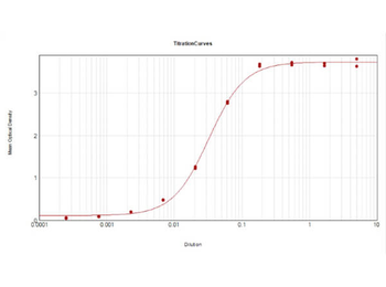
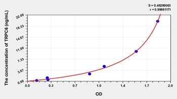
ELISA Results of Mouse Anti-TRPC6 Antibody. Each well was coated in duplicate with 0.1 µg of conjugate. The working dilution is 1:31000. The starting dilution of antibody was 5 µg/ml and the X-axis represents the Log10 of a 3-fold dilution. This titration is a 4-parameter curve fit where the IC50 is defined as the titer of the antibody. Assay performed using HRP conjugation Stabilizer, Rabbit Anti-Mouse IgG HRP conjugated (p/n orb347506) and TMB substrate (p/n orb348651).

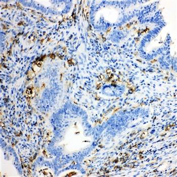
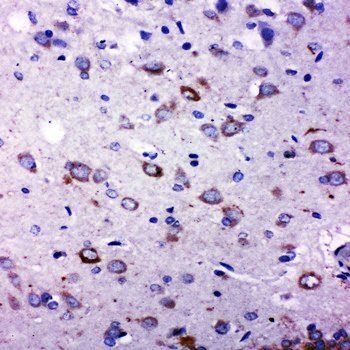
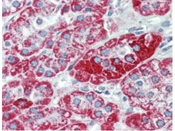
Immunohistochemistry using Biorbyt's anti-TRPC6 monoclonal antibody shows detection of TRPC6 in human adrenal (cortex) tissue (40X). The antibody was used a dilution to 2.5 µg/ml. The image shows strong staining with minimal background staining. Tissue was formalin fixed and paraffin embedded. No pre-treatment of sample was required. The image shows the localization of antibody as the precipitated red signal, with a hematoxylin purple nuclear counterstain.
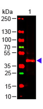
Western Blot of Mouse anti-TRPC6 Antibody. Lane 1: Mouse Kidney WCL (p/n orb348717). Load: 10 µg per lane. Primary antibody: TRPC6 Antibody at 1:1000 for overnight at 4°C. Secondary antibody: donkey anti-mouse DyLight™ 649 at 1:20000 for 30 min at RT. Block: orb348637 for 30 min at RT.

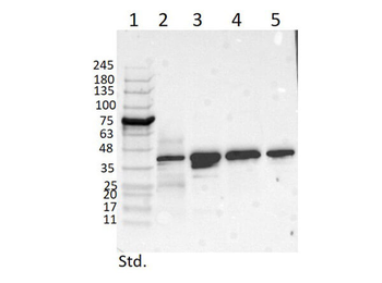
Western Blot of Mouse Anti-TRPC6 Antibody. Lane 1: Opal Prestained Molecular Weight Marker. Lane 2: Mouse Pancreas Tissue Lysate (p/n orb348719) [10 µg]. Lane 3: MCF-7 Whole Cell Lysate (p/n orb348664) [10 µg]. Lane 4: A431 Whole Cell Lysate (p/n orb348665) [10 µg]. Lane 5: Jurkat Whole Cell Lysate (p/n orb348674) [10 µg]. Primary Antibody: Anti-TRPC6 at 1 µg/ml overnight at 2-8°C. Secondary Antibody: Rabbit Anti-Mouse IgG Peroxidase (p/n orb347506) 1:40000 for 30 mins at RT. Blocking Buffer: BlockOut Buffer (p/n orb348644) for 30 mins at RT. Predicted MW: ~30kDa. Observed MW: ~40 kDa. Exposure: 5sec.
Documents Download
Datasheet
Product Information
Request a Document
Protocol Information
WB
Western Blot (IB, immunoblot)
IHC
Immunohistochemistry
ELISA
Enzyme-linked Immunosorbent Assay (EIA)
TRPC6 Antibody (orb344462)

- 0.0
Based on 0 reviews
Participating in our Biorbyt product reviews program enables you to support fellow scientists by sharing your firsthand experience with our products.
Login to Submit a ReviewAvailable Sizes
Select a size below
Choose Conjugation or Carrier Free Version
Free Secondary Antibody (20 ul)0/0
Please add an antibody product to your cart first.